
Page 266 - 2020_02-Haematologica-web
P. 266
S. Staessens et al.
staining can reveal novel molecular and cellular markers that could be extremely important for stroke pathophys- iology. The aim of this study was to assess and define the internal organization and common structural features of stroke thrombi, using specific immunohistochemical and immunofluorescence histology procedures.
Methods
Patient thrombi
Thrombi (n=188) were collected from acute ischemic stroke patients after a thrombectomy procedure was performed at the AZ Groeninge Hospital in Kortrijk, Belgium, regardless of prior treatment with rt-PA. All patients or their legal representative gave written consent under the approval of the AZ Groeninge Hospital ethical committee (AZGS2015065). Thrombi were retrieved using a stent retriever and/or aspiration device according to the judge- ment of the treating neuro-interventionalist. Thrombus material collected from multiple passes of one patient was pooled and fur- ther considered as one thrombus. Of the 188 collected thrombi, eleven thrombi were excluded because insufficient material was available to perform all analyses.
Thrombus histology
After retrieval, thrombi were gently removed from the device, washed in saline and immediately incubated in 4% paraformalde- hyde for 24 hours at room temperature. Next, samples were embedded in paraffin and cut into 5 μm sections. To check for dif- ferences in content throughout the thrombus, sections were ana- lyzed for fibrin, RBC, platelets, and von Willebrand Factor (vWF) every 75 μm in randomly selected thrombi. No substantial differ- ences in the quantity and general organization of these compo- nents were found between different sections of a single thrombus.12 Thus, one section per thrombus, exposing a large thrombus surface, was deemed representative and was used to quantify the Martius Scarlet Blue (MSB) staining.
Thrombus sections were stained with H&E (HT110216, Sigma- Aldrich, St. Louis, MO, USA), Martius Scarlet Blue [fibrin (dark pink/red) and RBC (yellow) staining] or Feulgen’s reaction [DNA staining (pink), 1079070001, Merck Chemicals, MA, USA]. Alternatively, thrombus sections were examined via immunohisto- chemistry and immunofluorescence for the presence of vWF (A008202-2, Dako, Glostrup, Denmark, and ab11713, Abcam, Cambridge, UK, respectively), platelets (GPIbα, MA5-11642, Invitrogen, Waltham, MA, USA), fibrin(ogen) (A0080, Dako), and leukocytes (CD45, 304002, Biolegend, San Diego, CA, USA). For
immunohistochemical stainings, nucleated cells were stained green using a Methyl Green solution (H-3402, Vector Laboratories). Images were acquired using a single slide scanner (Nanozoomer SQ, Hamamatsu Photonics, Japan). For immunofluorescent stain- ings, DNA was stained using 4,6-diamidino-2-phenylindole (DAPI, P36935, Invitrogen). RBC were visualized via their inherent autoflu- orescence at a wavelength of 555 nm. Images from immunofluores- cent stainings were acquired using an Axio Observer Z1 inverted fluorescent microscope (Zeiss, Carl Zeiss AG, Oberkochen, Germany) or a laser scanning confocal microscope (LSM710, Zeiss). Images were processed by Zen 2012 (blue edition, version 2.3, Zeiss) software. Negative controls of the immunohistochemical (Online Supplementary Figure S1A-D) and immunofluorescent (Online Supplementary Figure S1E and F) staining were achieved by omission of the primary antibody or by using isotype primary anti- bodies. A more detailed description of all histology procedures is provided in the Online Supplementary Methods.
Results
Red blood cell-rich and platelet-rich areas form distinct structural components of stroke thrombi
To better understand the structural features of acute ischemic stroke thrombi, we collected and histologically analyzed 177 thrombi retrieved by thrombectomy from patients with ischemic stroke. As shown in Online Supplementary Figure S2, the macroscopic appearance of retrieved thrombi was heterogenous in size, shape and color. All thrombi were sectioned and stained with H&E and MSB to visualize their general internal organization. H&E allows identification of fibrin/platelet aggregates (pink), RBC (red), and nucleated cells (dark blue) (Figure 1, left), whereas MSB staining selectively demonstrates the presence of fibrin (dark pink/red), RBC (yellow), and col- lagen (blue) (Figure 1, middle). H&E and MSB stainings revealed a typical common pattern in all thrombi, which is the presence of two distinct types of thrombus material: (i) RBC-rich/fibrin-poor material that appears red on H&E stainings and yellow on MSB stainings; and (ii) RBC- poor/fibrin-rich areas that appear as light pink areas on H&E staining and pink to red areas on MSB stainings. Interestingly, large amounts of blood platelets are only present in the RBC-poor/fibrin-rich areas and not in the RBC-rich areas, as shown via platelet-specific immunos- taining (Figure 1, right). Consequently, thrombi can be RBC-rich/platelet-poor (Figure 1A), mixed (Figure 1B), or
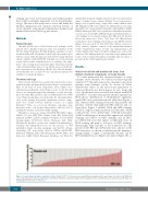
Figure 2. General stroke thrombus composition. Stroke thrombi (n=177, vertical bars) were quantitatively analyzed and the percentage of red blood cell (RBC)-rich areas (red) and platelet-rich areas (white) were determined. Thrombus composition ranges from very platelet-rich to very RBC-rich areas, with almost all thrombi con- taining significant amounts of both areas.
500
haematologica | 2020; 105(2)
