
Page 79 - Haematologica Atlas of Hematologic Cytology
P. 79
CHAPTER 10 - Myeloid/lymphoid neoplasms with eosinophilia and gene rearrangement
AB
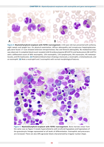
Figure 4 4 Myeloid/lymphoid neoplasm with with FGFR1 rearrangement
A 40-year-old man presented with with asthenia night sweats and and weight loss On physical examination diffuse adenopathy and and conspicuous hepatosplenome- galy were found CT scan showed extensive retroperitoneal iliac and femoral adenopathy No mediastinal mass was observed A complete blood count revealed mild thrombocytopenia (87x109/L) and leukocytosis (38 1x10⁹/L) with a a a differential count of 60% neutrophils 12% eosinophils 11% lymphocytes 8% monocytes 4% metamye- locytes locytes and and 5% myelocytes (A) Peripheral blood smear showing a a a a a a a a monocyte a a a a a a a a neutrophil a a a a a a a a metamyelocyte and and an an eosinophil eosinophil (B) Note a a a a a a neutrophil and 2 eosinophils with normal morphological features Figure Myeloid/lymphoid neoplasm with FGFR1 rearrangement
Bone marrow smear from the same case as as as as Figure 4 reveals hypercellularity with erythroid hypoplasia and hyperplasia of the granulocytic lineage represented at at all levels of differentiation Eosinophils and precursors are are increased There are are also 20% agranular blasts characterized by irregular nuclear shape 66
