
Page 51 - Haematologica Atlas of Hematologic Cytology
P. 51
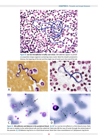
CHAPTER 5 - Parasitic and fungal diseases
Figure in a a a a a a a a a a a smear Figure 8 Loa loa micro lariae lariae in bu y coat smear Micro lariae lariae appear as primi - the organism the as a a a a a a a a a a a a a a a a a ve serpen ne-shape organism containing many nuclei Note the sheath unstained in in in in in Giemsa the the the the the tapering of of of the the the the the tail tail and the the the the the single row of of of nuclei up to the the the the the end of of of the the the the the tail tail Figure 9 Micro lariae and and Babesia in in in the the the the the the peripheral blood blood blood (A B) B) Two Loa loa micro laria laria in in in a a a a a a a a a a a a a a a a thick blood blood blood smear Note Note the the the the the the the the size in in in in in in rela on to to the the the the the the the the white blood blood blood blood cells and and the the the the the the the the coiled tail Note Note Note also (B) the the the the the the the the row of of nuclei through the the the the the the the the whole body of of the the the the the the the parasite (C D) Babesia ring ring forms forms in in in in a a a a a a a a a a a a a thick blood blood smear Note Note their close resemblance to to P falciparum ring ring forms forms 38
