
Page 36 - Haematologica Atlas of Hematologic Cytology
P. 36
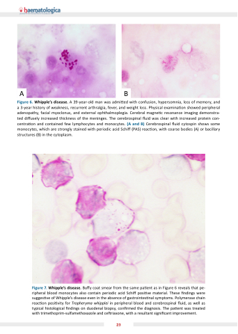
AB
Figure 6. Whipple’s disease. A 39-year-old man was admi ed with confusion, hypersomnia, loss of memory, and a 3-year history of weakness, recurrent arthralgia, fever, and weight loss. Physical examina on showed peripheral adenopathy, facial myoclonus, and external ophthalmoplegia. Cerebral magne c resonance imaging demonstra- ted di usely increased thickness of the meninges. The cerebrospinal  uid was clear with increased protein con- centra on and contained few lymphocytes and monocytes. (A and B) Cerebrospinal  uid cytospin shows some monocytes, which are strongly stained with periodic acid Schi  (PAS) reac on, with coarse bodies (A) or bacillary structures (B) in the cytoplasm.
Figure 7. Whipple’s disease. Bu y coat smear from the same pa ent as in Figure 6 reveals that pe- ripheral blood monocytes also contain periodic acid Schi  posi ve material. These  ndings were sugges ve of Whipple s disease even in the absence of gastrointes nal symptoms. Polymerase chain reac on posi vity for Tropheryma whipplei in peripheral blood and cerebrospinal  uid, as well as typical histological  ndings on duodenal biopsy, con rmed the diagnosis. The pa ent was treated with trimethoprim-sulfamethoxazole and ce riaxone, with a resultant signi cant improvement.
23
