
Page 276 - Haematologica Atlas of Hematologic Cytology
P. 276
ABAB
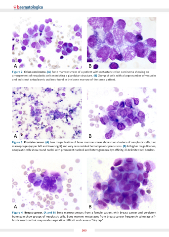
Figure 2 Colon carcinoma carcinoma (A) Bone marrow smear of a a a a a a a a a a a pa ent with metasta c c c c c c colon carcinoma carcinoma showing an arrangement of of of neoplas c c c c c c cells cells mimicking a a a a a a a a a glandular structure (B) Clump of of of cells cells with a a a a a a a a a large number of of of vacuoles and indis nct cytoplasmic outlines found in in in the the bone marrow of the the same pa ent AB Figure 3 Prostate cancer (A) Low magni ca ca on on of of bone marrow smear shows two two clusters of of neoplas c c c c c c cells two two macrophages (upper le and and lower right) and and very rare residual hematopoie c c c c precursors (B) At higher magni ca on neoplas c c c c c cells show round nuclei with prominent nucleoli and heterogeneous dye a a a nity ill-delimited cell cell borders AB Figure 4 Breast cancer cancer (A and and B) Bone marrow smears from a a a a a a a a a a a female pa ent ent with breast cancer cancer and and persistent bone pain show groups of neoplas c c c c cells Bone marrow metastases from breast cancer frequently s s s s s s s s s mulate a a a a a a a a a - bro c c c c reac on on that may render aspira on on di cult and cause a a a a a a a a a “dry tap” 263
