
Page 27 - Haematologica Atlas of Hematologic Cytology
P. 27
CHAPTER 2 - Cytological examination of bone marrow aspirate
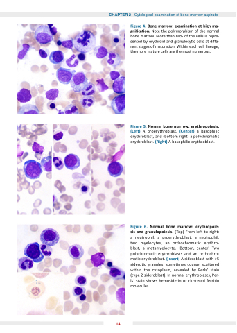
Figure ormal bone marrow erythropoiesis (Left) A proerythroblast (Center) a a a a a a a a a basophilic erythroblast erythroblast erythroblast erythroblast and (bottom right) a a a a a a a a a a polychromatic erythroblast erythroblast erythroblast (Right) A basophilic erythroblast erythroblast erythroblast Figure 4 one marrow examina on on on at high ma ma ma gni ca on on on on Note the the the polymorphism of of the the the normal bone marrow More than 80% of of the the the cells cells is is repre- sented by erythroid and granulocy c c c c c c cells cells at at di e- e- rent stages of matura on Within each cell cell cell lineage the the more mature cells are the the most numerous Figure ormal bone marrow erythropoie sis sis and granulopoiesis (Top) From left to right: a a a a neutrophil neutrophil a a a a proerythroblast a a a a neutrophil neutrophil two myelocytes an orthochromatic erythro- blast a a a metamyelocyte (Bottom center) Two polychromatic erythroblasts and an an orthochro- matic erythroblast (Insert) A sideroblast with >5 siderotic granules sometimes coarse scattered within the cytoplasm revealed by Perls’ stain (type 2 sideroblast) In normal erythroblasts Per- ls’ stain shows hemosiderin or clustered ferritin molecules 14
