
Page 26 - Haematologica Atlas of Hematologic Cytology
P. 26
AB
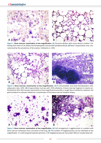
Figure 1 one marrow examina on on on on at low magni ca on on on on (A) Excessive dilu on on on on with sinous blood is evident Col- lec ng more than 0 5 mL dilutes the hematopoie c c c c marrow with peripheral blood (B) Note a a a a a a a a a hypocellular area cha- racterized by the prevalence of fat areolas Cellularity is <25% AB
Figure one one marrow marrow marrow examination at at at at at low magnification (A) A A A A normocellular area with with hematopoietic cells: adipocytes ratio >25% (B) A A A A hypercellular marrow marrow marrow with with >75% cellularity cellularity A A A A bone marrow marrow marrow fragment is is clearly visi- ble (bottom left) Microscopic examination examination at at at at at at low magnification provides a a a a a a a a a a a a a a a a a a a a a a a a rough idea of of cellularity cellularity cellularity however the the gold standard to to evaluate cellularity cellularity is examination examination of of the bone marrow trephine biopsy AB
Figure one marrow examina on on on at at low magni ca on on on (A) Cell arrangement in in in agglomerates in in in a a a a a a a a a a a a pa ent ent with bone marrow metastases from carcinoma of of the lung (B) The number of of megakaryocytes can be be es es es mated at at low magni ca on In a a a a a a a a a a well-prepared aspirate
specimen 2-10 megakaryocytes per low-power eld are are usually observed 13
