
Page 21 - Haematologica Atlas of Hematologic Cytology
P. 21
CHAPTER 1- Morphological examination of peripheral blood
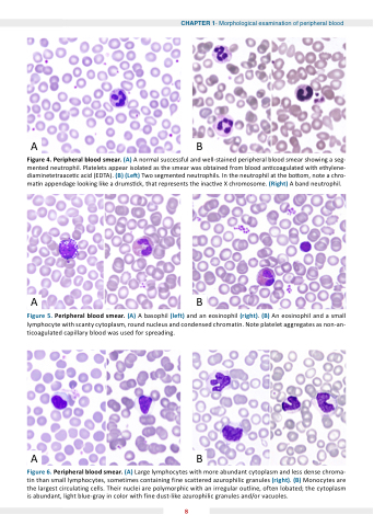
Figure 4 Peripheral blood
blood
blood
smear smear smear (A) A A normal successful and well-stained peripheral blood
blood
blood
smear smear smear showing a a a a a a a a a a a a a a a a a a a seg- mented mented neutrophil neutrophil neutrophil Platelets appear isolated as as the the the smear smear smear was obtained from blood
blood
blood
an an coagulated with ethylene- diaminetetraace c c c c c c c c acid (EDTA) (B) (Le ) ) ) ) Two segmented neutrophils In the the the the neutrophil neutrophil neutrophil neutrophil at at at at at the the the the bo om om om om note a a a a a a a a a a a a a a a a a a a a a a a a chro- ma n n n n n n n n n n n n n appendage looking like a a a a a a a a a a a a a drums ck that represents the the the inac ve X chromosome (Right) A A band neutrophil neutrophil neutrophil Figure Peripheral blood
smear (A) and and (B) Figure 5 Peripheral blood
smear (A) A A A basophil (left) and and an an an eosinophil eosinophil (right) (B) An eosinophil eosinophil and and a a a a a a a a small can on on lymphocyte with scanty cytoplasm round nucleus and and and condensed chromatin Note platelet aggregates as as as non-an-
ticoagulated capillary blood
was used for spreading Figure 6 Peripheral blood
smear (A) Large lymphocytes lymphocytes with more abundant cytoplasm and less dense chroma- tin tin than small lymphocytes lymphocytes sometimes containing fine scattered azurophilic granules (right) (B) Monocytes are are the the largest circulating cells Their nuclei are are polymorphic with with an an an an an an irregular outline often lobated the the cytoplasm is abundant light blue-gray in in in in color with with fine dust-like azurophilic granules and/or vacuoles 8
