
Page 17 - Haematologica Atlas of Hematologic Cytology
P. 17
CHAPTER 1 - Morphological examination of peripheral blood
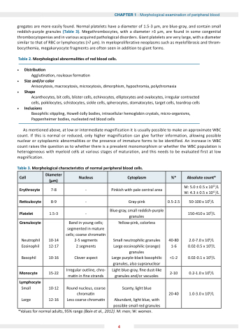
gregates are are more easily found Normal platelets have a a a a a a a a a a a a a diameter of 1 5-3 μm are are blue-gray and contain small reddish-purple granules (Table 3) Megathrombocytes with a a a a a a a diameter >3 μm are found in some congenital thrombocytopenias and in various acquired pathological disorders Giant platelets are very large with a a a a a a a a a a a a diameter similar to that of of RBC or lymphocytes (>7 μm) In myeloproliferative neoplasms such as as myelofibrosis and throm- bocythemia megakaryocyte fragments are often seen in addition to giant forms Table 2 Morphological abnormali es of red blood
cells Distribu on Agglu na on on rouleaux forma on on Size and/or color
Anisocytosis macrocytosis microcytosis dimorphism hypochromia polychromasia Shape
Acanthocytes bit cells cells blister cells cells echinocytes elliptocytes and ovalocytes irregular contracted cells cells cells cells poikilocytes schistocytes sickle cells cells cells cells spherocytes stomatocytes target cells cells cells cells teardrop cells cells cells cells Inclusions
Basophilic s s s s s s s ppling Howell-Jolly bodies intracellular hemoglobin crystals micro-organisms Pappenheimer bodies nucleated red blood
cells As mentioned above at at at at low or intermediate magnification it is usually possible to make an approximate WBC count If this is is normal or or or reduced only higher magnification can give further information allowing possible nuclear or or or or cytoplasmic abnormalities or or or or the presence of immature forms to to be identified An increase in in WBC count raises the the the the the question as to whether whether there is is is is a a a a a prevalent monomorphism or or whether whether the the the the the WBC population is is is is heterogeneous with myeloid cells at at at at at various stages of maturation and this needs to be evaluated first at at at at at low magnification Table 3 Morphological characteris cs of normal peripheral blood
cells Cell Diameter (μm)
Nucleus
Cytoplasm
%*
Absolute count*
Erythrocyte
7-8 - Pinkish with pale central area M: 5 5 0 0 0 ± 0 0 0 5 5 x 10¹²/L W: 4 3 ± 0 0 5 x 10¹²/L Re culocyte
8-9
- Gray-pink
0 5-2 5 5 50-100 x 109/L
Platelet
1 5-3 - Blue-gray small reddish-purple granules 150-410 x 109/L
Granulocyte
Neutrophil Eosinophil
Basophil 10-14 12-17
10-16
Band in young cells segmented in mature cells coarse chroma n 2-5 segments
2 segments
Clover aspect
Yellow-pink colorless
Small neutrophilic granules Large eosinophilic (orange) granules Large purple-black basophilic granules also supranuclear
40-80 1-6
<1-2
2 0-7 0 0 0 x 109/L
0 0 0 0 02-0 5 x 10⁹/L
0 0 0 0 02-0 1 1 x 10⁹/L
Monocyte
15-22
Irregular outline chro- ma n n n n in ne strands
Light blue-gray ne dust-like granules and/or vacuoles
2-10
0 0 0 2-1 0 0 0 x 109/L
Lymphocyte
Small Large 10-12 12-16
Round nucleus coarse chroma n Less coarse chroma n Scanty light blue Abundant light blue with possible small red granules 20-40
1 1 0-3 0 0 0 x 109/L
*Values for normal adults 95% range (Bain et al al al 2012) M: men men W: women 4
