
Page 16 - Haematologica Atlas of Hematologic Cytology
P. 16
Chapter 1 MORPHOLOGICAL EXAMINATION OF PERIPHERAL BLOOD
Morphological examination of the the peripheral blood
(PB) still represents a a a a a a a basic step in in the the hematologic work- up and is is is essential for for a a a a a a a a correct diagnosis An expert cytologist can certainly acquire more information from this than from any other single test Since nowadays automated analyzers are able to to provide some of the the same information criteria have been proposed that establish the the the indications for for the the the assessment of the the the PB smear The need for PB film review is indicated by qualitative flags and/or quantitative abnormal results of the complete blood
blood
blood
count (Barnes et et al al al 2005 Palmer et et al al al 2015) Non-anticoagulated venous or or capillary blood
blood
blood
or or blood
blood
blood
anticoagulated with ethylenediaminetetraacetic acid (EDTA) can be used for spreading In the first case possible artifacts due to to to the the the storage and the the the effects of of anticoagulants as as well as as the the the possibility of of pseudothrombocyto- penia and pseudoleukocytosis are avoided The smear should be quickly air-dried as prolonged drying can cause morphological alterations especially in in the the red blood
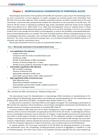
cells (RBC) Another possible artifact of spreading is cell cell distortion The smear is is then stained with panoptic dyes as in in the the May-Grünwald Giemsa (MGG) method or or if indicated with specific cytochemical stains Microscopic examination must be carried out systematically (Table 1) Table 1 Microscopic examina on of the peripheral blood
smear Low magni ca on (10x objec ve) Quality of the smear Comparison with the results of complete blood
count Distribu on of RBC Number and and distribu on of WBC and and platelets Presence of abnormal large cells i e e e e e e blasts
Iden fy an area suitable for examina on on at higher magni ca on on Intermediate magni ca on on (40x objec ve) RBC and WBC morphology
Presence of abnormal cells Approximate es ma ma on of WBC count (mean WBC WBC count count per eld x 2 000 = WBC WBC count/mm3)*
High magni ca on on (100x oil immersion lens) WBC di eren al count Approximate es ma ma on of platelet count (mean platelet platelet count count per eld x 20 000 = platelet platelet count/mm3)*
Detec on of cytological details
RBC: red blood
blood
cell cell WBC: white blood
blood
cell cell *Maedel and Doig 2012 The International Council for Standardization Standardization in Haematology (ICSH) Committee on on on on Standardization Standardization of Pe- ripheral Blood Cell Morphology Nomenclature and and Grading has provided guidelines for the nomenclature and and grading of RBC white blood
cell (WBC) and platelet abnormalities (Palmer et et al al al 2015) An ideal area for evaluation is that where RBC are are only slightly overlapped The morphological examination of RBC includes the the the evaluation of their distribution distribution size and and shape hemoglobin concentration and and distribution distribution the the the possible presence of inclusions and parasites or nucleated RBC (Table 2) In normal conditions RBC shape shape and size are rather homogeneous: round or or slightly oval in in shape shape 7-8 μm in in diameter with an area of of central pallor occupying about one-third of of the cell volume In pathological contitions various morphological alterations may be observed (i) Changes in shape (poikilocytosis) and size (anisocytosis) may reflect a a a a a a a broad range of of of disorders of of of erythropoiesis (ii) The inadequate synthesis of of of hemoglobin leads to changes in in in staining: hypochromia hyperchromia dimorphic pattern (iii) Peripheral RBC damage may cause sphe- rocytosis fragmentation whereas attempts by marrow to to compensate anemia cause stimulation of erythropoie- sis with the consequent appearance of of signs of of immaturity such as as as polychromasia and erythroblastemia As regards platelets the the the microscopic examination of the the the PB allows the the the following parameters to be evalua- ted: approximate count size morphology
including granularity presence of aggregates or or satellitism In case of thrombocytopenia the the entire smear needs to to be evaluated carefully especially the the tails
where platelet ag-
3
