
Page 169 - Haematologica Atlas of Hematologic Cytology
P. 169
CHAPTER 17 - - Mature B-cell neoplasms
AB
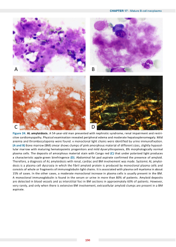
CD Figure 24 AL amyloidosis A A 54-year-old man presented with nephrotic syndrome renal impairment and restri- ctive cardiomyopathy Physical examination revealed peripheral edema and moderate hepatosplenomegaly Mild anemia and thrombocytopenia were were found monoclonal light chains were were identified by urine immunofixation (A and B) Bone marrow (BM) smear shows clumps of of pink amorphous material of of different sizes slightly hypocel- lular marrow with maturing hematopoietic progenitors and mild dyserythropoiesis 8% morphologically normal plasma cells The deposits of amorphous material stain with Congo red (C) that under polarized light produces a a a a a a a a a a characteristic apple-green birefringence (D) Abdominal fat pad aspirate confirmed the presence of amyloid Therefore a a a a a a a a a a diagnosis of AL AL amyloidosis with renal cardiac and BM involvement was made Systemic AL AL amyloi- dosis is is is a a a a a a a a a a plasma plasma cell cell dyscrasia in in which the fibril amyloid protein is is is produced by monoclonal plasma plasma cells and consists of of whole or fragments of of immunoglobulin light chains It is is associated with plasma cell myeloma in in in about 15% of cases cases In the the the other cases cases a a a a a a a a a moderate monoclonal increase in in in plasma cells is usually present in in in the the the BM A A monoclonal immunoglobulin is found in in in in the serum or or urine in in in in more than 80% of patients Amyloid deposits are detected in in in in blood vessels and as interstitial foci in in in in BM sections in in in in approximately 60% of patients However very rarely and only when there is extensive BM BM involvement extracellular amyloid clumps are are present in in a a a a a a a BM BM aspirate 156
