
Page 168 - Haematologica Atlas of Hematologic Cytology
P. 168
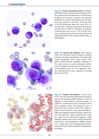
Figure 21 Plasma cell leukemia (PCL) Peripheral blood (PB) smear showing immature plasma cells Rare plasma cells are are observed in the PB smears of of of about 15% of of of cases of of of plasma cell myeloma (PCM) PCL is a a a a PCM PCM in which plasma cells are gre- ater than than 2 2 0x109/L in the PB or or more than than 20% in the PB differential white cell count PCL is a a rare disorder: it may be primary and represent an an unusual onset of PCM or secondary as leukemic transformation that occurs in 1% of PCM cases The circulating plasma cells may have features of lymphoplasmacytic cells cells typical plasma plasma cells cells or plasmablasts Figure 22 Plasma cell leukemia Bone marrow smear from the same case as as in Figure 21 Diffuse infiltration by large plasmablasts with rather high nuclear:cytoplasmic ratio loose nuclear nuclear chro- matin evident nucleoli basophilic cytoplasm In PCL bone marrow is usually extensively infiltra- ted and malignant plasma cells are found also in extramedullary sites PCL is is an aggressive dise- ase characterized by poor response to therapy and short survival Figure 23 Plasma cell leukemia In some cases of PCL the cells show blast-like features and poor signs of morphological differentiation so that the diagnosis is is is often difficult Immunocytology is is is useful for identification and to distinguish from acute leukemia blasts and lymphoma cells It should be noted however that in PCL CD20 an- tigen is more frequently expressed and CD56 an- tigen less frequently expressed than in other pla- sma cell myelomas Some cytoenzymatic stains are useful for the differential diagnosis (Left) Leukemic plasma cells show strong paranuclear reactivity for acid phosphatase (Right) Cytopla- smic light chains are demonstrated by immu- nocytochemistry (immuno-alkaline phosphatase technique) 155
