
Page 102 - Haematologica Atlas of Hematologic Cytology
P. 102
ABAB
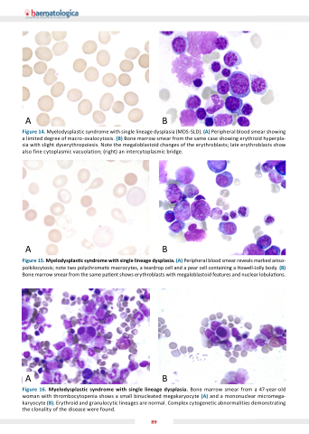
Figure 14 Myelodysplastic syndrome with single lineage dysplasia (MDS-SLD) (A) Peripheral blood smear smear showing showing a a a a a a a a a a a a a a a a a a a a a limited degree of of macro-ovalocytosis (B) Bone marrow smear smear from the the the same case showing showing erythroid hyperpla- sia with slight dyserythropoiesis Note the the the megaloblastoid changes of of the the the erythroblasts erythroblasts late erythroblasts erythroblasts show show also fine cytoplasmic cytoplasmic vacuolation (right) an an intercytoplasmic bridge AB Figure 1 Myelodysplas c syndrome with single lineage dysplasia (A) Peripheral blood smear reveals marked aniso- poikilocytosis note two polychroma c c c c c c c c macrocytes a a a a a a a a a teardrop cell cell and a a a a a a a a a pear cell cell containing a a a a a a a a a Howell-Jolly body (B) Bone marrow smear from the same pa ent shows erythroblasts with megaloblastoid features and nuclear lobula ons AB Figure 1 Myelodysplastic syndrome with with single lineage dysplasia Bone marrow smear from a a a a a a a a a a a a a a a a a a a 47-year-old woman with with thrombocytopenia shows a a a a a a a a a a a a a a a a a a a a a a a a a a a a small binucleated megakaryocyte (A) and and a a a a a a a a a a a a a a a a a a a a a a a a a a a a mononuclear micromega- karyocyte karyocyte (B) Erythroid and and granulocytic lineages are normal normal Complex cytogenetic abnormalities demonstrating the the clonality of the the disease were found 89
