
Page 101 - Haematologica Atlas of Hematologic Cytology
P. 101
CHAPTER 12 - Myelodysplastic syndromes
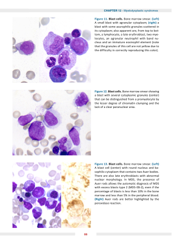
Figure 11 Blast cells Bone marrow smear (Left) A small blast with agranular cytoplasm (right) a a a a a a a blast with some azurophilic granules scattered in its cytoplasm also apparent are are from top top to to to bot- tom a a a a lymphocyte a a a a late erythroblast two mye- locytes an an an agranular neutrophil with band nu- cleus and an an immature eosinophil element (note that the granules of this cell are not yellow due to the difficulty in in correctly reproducing this color) Figure 1 Blast cells Bone marrow smear showing a a a a a blast with several cytoplasmic granules (center) that can be dis nguished from a a a promyelocyte by the the lesser degree of chroma n n n clumping and the the lack of a a a a a a a a clear clear paranuclear area Figure 1 Blast cells Bone marrow smear (Left) A blast cell (center) with round nucleus and ba- sophilic cytoplasm that contains two Auer bodies There are also late erythroblasts with abnormal nuclear morphology In MDS the presence of Auer rods allows the automatic diagnosis of MDS with excess blasts type 2 2 (MDS-EB-2) even if the percentage of blasts is less than 10% in the bone marrow and less than 5% in the peripheral blood (Right) Auer rods are better highlighted by the peroxidase reaction 88
