
Page 75 - 2019_12-Haematologica-web
P. 75
Myeloperoxidase in myelodysplastic syndromes
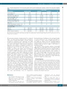
Table 4. Baseline characteristics for 68 consecutive patients with suspected myelodysplastic syndromes enrolled in the prospective validation study.
Confirmed MDS
Characteristics*
Female gender, n (%)
Age, mean (SD), y
Hemoglobin, median (IQR), g/dL
Platelet, median (IQR), ×109/L
ANC, median (IQR), ×109/L
All patients (N=68)
No (N=53)
22 (42)
73.6 (9.2) 10.3 (9.6–12.4) 124 (72–205)
3.2 (2.3–4.9)
Yes (N=15) P (47) 0.72
Creatinine, median (IQR), μmol/L
29
74.7 10.4 119 3.4 92 29/39
(43)
(9.2) (9.6–12.6) (80–198) (2.1–4.9) (73–114) (74)
93 (76–116) 24/33 (73)
7
78.4 10.7 104 3.8 83 5/6
(8.4) 0.07 (9.6–14.1) 0.56 (80–148) 0.77 (1.8–5.3) 0.69 (69–99) 0.22 (83) 0.99
C-reactive protein ≥ 3 mg/L, n (%)
8 (12) 8 (15) - (-) -
ICUS,n(%)
Confirmed myelodysplastic syndrome, n (%)
Neutrophil MPO expression in peripheral
blood, median (IQR) Mean, FI
Median, FI
Robust coefficient of variation, %
15
4040 3883 31.9
(22)
(2828–5739) (2730–5500) (29.5–34.6)
- (-) 15 3981 (2816–5292) 4296
3816 (2732–5184) 4175
31.0 (28.9–32.5) 38.1
(100) - (2840–6362) 0.46
(2701–6167) 0.61
N/n: number; ANC: absolute neutrophil count; FI: fluorescence intensity; ICUS: idiopathic cytopenia of undetermined significance; IQR: interquartile range (25–75th percentiles); MDS: myelodysplastic syndrome; MPO: myeloperoxidase; N/n: number; SD: standard deviation. *Values were missing for platelet count (n=2), C-reactive protein (n=29), and creatinine (n=25) concentrations.
68 consecutive patients routinely referred for suspected MDS. 2) Control subjects included in the retrospective study did not undergo BM aspirate or biopsy, with the potential for verification bias.38 Although overt MDS could not be formally excluded in these subjects, none of the controls had evidence of PB cytopenia, making this hypothesis very unlikely. 3) Peripheral cytopenia was defined based on standard laboratory values, as recom- mended by others.18,23 To assess the robustness of our findings, we repeated the analysis after restricting the study sample to patients with evidence of cytopenia according to WHO categorization, and the diagnostic accuracy estimates were similar although less precise (Online Supplementary Table S10). 4) Neutrophils of MDS patients can exhibit varying levels of CD14, CD64, or CD16 expression compared with healthy controls. However, we did not have any difficulty separating neu- trophils from monocytes because of increased CD14 expression. CD64 was not used in the gating strategy and any modulation of its expression would not alter the results. We rarely observed downmodulation of CD16 in this series and these cells were infrequent among the granulocyte population. Importantly, the RCV for MPO expression of circulating neutrophils remained unchanged depending on whether or not these cells were taken into account. 5) The diagnosis of MDS can be deli- cate with subtle cytological signs of myelodysplasia. There is some evidence that cytomorphology examina-
hematopathologists. Furthermore, the cytological dyspla- sia criterion threshold of 10% abnormal cells limited to one lineage is a subject of debate. 6) Our diagnostic accu- racy study was carried out in two university-affiliated hospitals in France. For this reason, our findings may lack external validity and may not apply to other regions or healthcare settings.
In conclusion, flow cytometric analysis of neutrophil MPO expression in PB might increase the diagnostic yield of BM aspirate in patients referred for suspected MDS. A RCV value <30.0% accurately rules out MDS, with both sensitivity and negative predictive value estimates of 100%. This strategy might obviate the need for invasive BM aspirate for up to 29% of patients with suspected MDS in real-life practice. Although promising, these pre- liminary results require replication in a large multicenter prospective diagnostic accuracy study.
Acknowledgments
Becton Dickinson Bioscience provided antibodies free of charge. This research received no other specific grant from any funding agency in the public, commercial, or non-profit sectors. Statistical analysis was performed within the Grenoble Alpes Data Institute (ANR-15-IDEX-02). The authors thank Séverine Beatrix, Laure Chevrolat, Ghislaine Del-Vecchio, Richard Di Schiena, Michel Drouin, Claire Guillier, Frédérique Martinez, and Christine Vallet for their technical assistance. The authors also thank Linda Northrup, English Solutions (Voiron, France) for her assistance in preparing and editing the manuscript.
tion lacks reproducibility, even
References
1. Arber DA, Orazi A, Hasserjian R, et al. The 2016 revision to the World Health Organization classification of myeloid neo- plasms and acute leukemia. Blood. 2016;127(20):2391-2405.
2. Fenaux P, Haase D, Sanz GF, et al. Myelodysplastic syndromes: ESMO Clinical
for experienced
Practice Guidelines for diagnosis, treatment and follow-up. Ann Oncol. 2014;25(Suppl 3):iii57-69.
3. Tefferi A, Vardiman JW. Myelodysplastic syndromes. N Engl J Med. 2009;361 (19):1872-1885.
4. Malcovati L, Hellstrom-Lindberg E, Bowen D, et al. Diagnosis and treatment of primary myelodysplastic syndromes in adults: rec-
ommendations from the European LeukemiaNet. Blood. 2013;122(17):2943- 2964.
5. Gangat N, Patnaik MM, Tefferi A. Myelodysplastic syndromes: Contemporary review and how we treat. Am J Hematol. 2016;91(1):76-89.
6. Buckstein R, Jang K, Friedlich J, et al. Estimating the prevalence of myelodysplas-
(32.7–50.2)
<0.001
haematologica | 2019; 104(12)
2389
