
Page 201 - 2019_12-Haematologica-web
P. 201
Antithrombin deficiency and pediatric thrombosis
sis-free survival between males and females are also illus- trated by Kaplan-Meier survival curves (Figure 3).
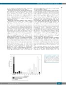
Analysis by age revealed two periods with higher preva- lence of thrombosis: adolescence (n=49, 67.1%) and the neonatal period (n=15, 20.5%) (Figure 2). In adolescents, clinical presentation was similar to adults: deep vein thrombosis of lower limbs and/or pulmonary embolism (n=47). They also shared the same predisposing factors as adults: pregnancy/puerperium, oral contraceptive use, trauma, immobilization or surgery (n=22) (Online Supplementary Table S1).
Remarkably, in neonates, thrombosis often occurred at unusual sites (11 of 15, 73.3%) (Table 1) such as upper extremities, renal veins and cerebral veins. Four patients suffered from arterial thrombosis, with associated venous thrombosis in two of them. In seven neonates, the throm- botic events were idiopathic while in the other eight, pos- sible provoking factors were identified: complicated deliv- ery (forceps or vacuum extraction), infection/sepsis, trau- ma, surgery or fetal distress (Figure 2 and Online Supplementary Table S1). Only one thrombotic event was associated with the presence of a central venous catheter.
The prevalence of cerebral sinovenous thrombosis (CSVT) was very high in our cohort (n=13; 17.8%), espe- cially at a young age (8 neonates and 4 children <6 years) (Figure 2 and Online Supplementary Table S1). It is notewor- thy that in three cases CSVT occurred after assisted deliv- ery (emergency caesarian section, forceps or vacuum extraction). It is interesting to note the extreme severity of the events. Six children (8.2%) died as a consequence of a thrombotic episode. If we only consider neonatal throm- bosis, fatality rate rises to 20% (3 of 15) (Online Supplementary Table S1). Interestingly, two of the deceased neonates were unrelated homozygous carriers of the p.Leu131Phe variant, responsible for Antithrombin Budapest III, a type II heparin binding site deficiency.14 Moreover, morbidity after pediatric thrombosis was severe. One child needed to have an arm amputated,
another developed serious psychomotor retardation, and one had permanent tetraplegia.
From a molecular/biochemical point of view, the symp- tomatic children in our study predominantly showed type I antithrombin deficiency (76.7%). This results in a signif- icantly higher thrombotic risk associated with type I defi- ciency compared to type II, with an odds ratio of 2.3 (95%CI: 1.26-4.18; P=0.007). Only 14 patients carried a type II deficiency: six were type II RS or PE and eight type II HBS deficiency (Online Supplementary Table S1). In patients with type I deficiency, around half of the throm- botic events were unprovoked while this was much high- er (75%) among patients with type II HBS deficiency. In most patients (6 of 8) with type II HBS deficiency the p.Leu131Phe variation was detected; four were homozy- gous and the two heterozygous cases were also carriers of the Factor V Leiden mutation (one heterozygous and one homozygous) (Online Supplementary Table S1). Considering the whole cohort of subjects with antithrom- bin deficiency, only eight out of 223 subjects with type II HBS deficiency (3.6%) suffered from thrombosis during childhood and, as indicated before, most of them carried additional genetic risk factors or had the SERPINC1 muta- tion in homozygous state. The prevalence of pediatric thrombosis in the whole cohort of individuals with type I deficiency was higher: 56 out of 604 (9.3%).
In two patients, the molecular mechanism responsible for the antithrombin deficiency was not found. These patients showed low anti-FXa activity on several inde- pendent blood samples and had first degree family mem- bers with the same low antithrombin values. One patient had a congenital disorder of N-glycosylation as the under- lying cause of the deficiency (Online Supplementary Table S1).19
The p.Leu131Phe mutation was the most prevalent mutation in our pediatric cohort with six carriers belong- ing to five families. Four unrelated patients carried the c.1154-14G>A mutation affecting splicing and four
Figure 2. Distribution of thrombotic events among children with antithrombin deficiency according to age. Localization of the thrombo- sis is also represented: deep vein thrombosis (DVT) of the lower limbs and/or pulmonary embolism (PE) (white), cerebral sinovenous thrombosis (black), or unusual localizations (gray).
haematologica | 2019; 104(12)
2515
