
Page 148 - 2019_12-Haematologica-web
P. 148
F. Späth et al.
AB
CD
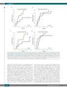
Figure 4. Probability of progression of monoclonal gammopathy of undetermined significance to multiple myeloma depending on transforming growth factor-alpha level at pre-diagnostic repeated sampling. Patients with low transforming growth factor-alpha (TGF-a) levels are represented in green, those with high levels in blue. Probability of progression of monoclonal gammopathy of undetermined significance (MGUS) according to TGF-a levels in patients with (A) low- (no risk factor) or low- intermediate-risk (one risk factor) MGUS (n=28) or (B) high-intermediate- (two risk factors) or high-risk (three risk factors) MGUS (n=22). Risk factors considered were: M-protein ≥15 g/L, non-IgG MGUS, and abnormal free light chain (FLC) ratio.4 Risk of progression of MGUS according to TGF-a levels in patients with (C) stable (n=16) or (D) increasing M-protein levels or involved FLC ratios (n=25) between baseline and repeated samples. Increasing M-protein levels and involved FLC ratios were defined by an increase ≥25% with either an absolute rise ≥5 g/L for M-protein levels or ≥100 mg/L for involved light chains.
and is involved in the regulation of lymphangiogenesis.28 Furthermore, serum levels of FGF-2 were found to be lower in individuals with systemic sclerosis than in healthy controls, possibly reflecting underlying defective angiogenesis in the former.29 This could be of interest as systemic sclerosis is associated with an increased risk of developing B-cell lymphoid malignancies.30 TGF-a, a lig- and of the epidermal growth factor receptor, is a mediator of oncogenesis and malignant progression.31 It is thus bio- logically counterintuitive that we observed decreasing plasma levels of TGF-a among future myeloma cases. Dysregulated blood marker levels could theoretically reflect cancer immune evasion32 or be a result of the ongo- ing disease process including a shift of immune-related cells towards the bone marrow microenvironment.33 However, to the best of our knowledge these processes
have not been reported to involve TGF-a. Our data indi- cated that the levels of VEGF, FGF-2, and TGF-a were higher in cases than in controls, decades before their diag- nosis of MM (all P>0.05). Interestingly, it was recently shown that blood levels of VEGF and TGF-a are largely influenced by heritable factors.34 One might speculate that this could reflect a genetic predisposition, leading to reversed plasma levels of these proteins during the ongo- ing disease process. It is important to note that plasma marker levels do not necessarily reflect microenvironmen- tal conditions in the bone marrow. This was illustrated by a small study, including 30 MM patients and 10 healthy controls, in which higher TGF-a levels were observed in the bone marrow of MM patients than in controls, but lev- els in the peripheral blood were lower in the MM patients than in the controls, although the differences did not reach
2462
haematologica | 2019; 104(12)
