
Page 108 - 2019_12-Haematologica-web
P. 108
M. Wu et al.
AD
B
C
E
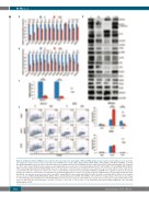
Figure 2. DOCK2 knockdown in MV4;11 cells resulted in decreased expression and activity of FLT3 and DNA damage response factors. (A) Quantitative reverse transcrip- tase polymerase chain reaction assays revealed decreased mRNA levels of FLT3, CHK1, WEE1, MSH2, MSH6, MLH1, PIM-1, RAD51, JUN, MYB and MEIS1 in DOCK2 knockdown (KD) MV4;11 cells. The levels of the transcripts were normalized based on that of GAPDH, and the relative expression of each transcript in KD cells compared to control cells is shown. (B) Western blot analysis revealed significantly decreased levels of total and phosphorylated FLT3, CHK1, WEE1, JUN, total MSH2, MLH1, RAD51, PIM-1, and phosphorylated histone H2AX (gH2AX). The level of expression of each protein was normalized to the expression level of β-actin, and the relative expression of each protein in KD cells compared to control cells is shown. (C) DOCK2 KD resulted in decreased binding of MEIS1/2 and MYB to the regulatory element located -15 kb from the FLT3 initiation codon. Relative enrichments were normalized against those in control cells. (D) The reduction in DNA damage response (DDR) activity in DOCK2 KD MV4;11 cells was due to the decrease in Rac1 and FLT3 activity. MV4;11 cells treated with NSC23766 (NSC; 40 μM) or sorafenib (SB; 25 nM) for 20 h exhibited decreased levels of MEIS1, MYB, MSH2, MLH1, RAD51, PIM-1, and phosphorylation of STAT5, CHK1, WEE1, JUN and FOS. (D) Compared with control cells, the percentage of cells harboring elevated gH2AX levels in DOCK2 KD MV4;11 cells was increased upon treatment with ara-C (3 μM) and decreased upon treatment with 5-fluorouracil (5-FU; 0.5 μM). Cells were treated for 18 h. *P<0.05; **P<0.01; ***P<0.001; ****P<0.0001. C: cells expressing control short hairpin (sh)RNA; KD: cells expressing shRNA against DOCK2.
2422
haematologica | 2019; 104(12)
