
Page 131 - 2019_11 Resto del Mondo-web
P. 131
CUDC-907 monotherapy in AML
Supplementary Figure S7). DNA strand breaks induced by CUDC-907 were also detected in two primary AML patient samples but not in normal human bone marrow mononuclear cells (Figure 6E), suggesting that CUDC-907 does not induce DNA damage in normal hematopoietic cells. To determine the functional role of CHK1, Wee1, and RRM1 in apoptosis induced by CUDC-907, U937 cells were treated with CUDC-907 alone or in combina- tion with the CHK1 inhibitor LY2603618, Wee1 inhibitor MK-1775, or the RR inhibitor hydroxyurea for 24 h. Annexin V/PI staining and flow cytometry revealed that each inhibitor significantly enhanced CUDC-907-induced apoptosis (Figure 6F), which suggests that CHK1, Wee1, and RRM1 also play important roles in CUDC-907- induced apoptosis in the cells. Real-time reverse transcrip- tase PCR results showed that CUDC-907 treatment caused significant decreases of CHK1, Wee1, and RRM1 transcripts in the AML cells both in vitro and in vivo (Figure 6G-J), suggesting that CUDC-907 downregulates CHK1, Wee1, and RRM1 expression in the cells through tran- scriptional regulation. While it has been reported that non- isoform selective PI3K inhibitors also inhibit DNA-PK, inhibition of DNA-PK is not likely to have contributed to the increased DNA damage-induced by CUDC-907 since its effect on DNA-PK activity was minimal (Online Supplementary Figure S8).
CUDC-907 downregulates c-Myc in acute myeloid leukemia cells
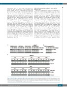
CUDC-907 treatment was shown to downregulate c- Myc protein in diffuse large B-cell lymphoma cells.16 Since c-Myc is an oncoprotein that is frequently activated in AML cells and plays a role in leukemogenesis,29,30 we next determined the role of c-Myc in CUDC-907-induced apoptosis in AML cells. Indeed, CUDC-907 treatment decreased expression of c-Myc in AML cell lines and a pri- mary AML sample (Figure 7A). In addition, decreased expression was detected in our MV4-11 xenograft mouse model following a single dose of CUDC-907 (Figure 7B). The pan-caspase inhibitor Z-VAD-FMK did not have an effect on downregulation of c-Myc by CUDC-907 (Online Supplementary Figure S9A). Furthermore, treatment with SAHA, GDC-0941, and SAHA plus GDC-0941 did not reduce c-Myc protein levels, again suggesting that the hybrid is more potent than the parental compounds (Online Supplementary Figure S9B). In both MOLM-13 and U937 cells, downregulation of c-Myc was detected as early as 4 h after CUDC-907 treatment (Figure 7C). c-Myc transcript levels were decreased in AML cell lines, two pri- mary AML patient samples (Figure 7D) and in the MV4-11 xenograft mouse model following a single dose of CUDC- 907 (Figure 7E). Overexpression of c-Myc resulted in par- tial inhibition of CUDC-907-induced apoptosis (Figure
AB
C
Figure 7. CUDC-907 treatment downregulates c-Myc in acute myeloid leukemia cells. (A) Acute myeloid leukemia (AML) cell lines and primary AML patient sample AML#77 were treated with CUDC-907 for 24 h. Whole cell lysates were subjected to western blotting. Normalized densitometry measurements are shown. (B) the MV4-11 xenograft model was treated with a single dose of CUDC-907 21 days after cell injection. Bone marrow cells were harvested 24 h after treatment. Human cells were enriched and then whole cell lysates were subjected to western blotting. (C) MOLM-13 and U937 cells were treated with CUDC-907 for up to 24 h. Whole cell lysates were subjected to western blotting. (continued on the next page)
haematologica | 2019; 104(11)
2237
