
Page 182 - 2019_10 resto del Mondo_web
P. 182
S. Bhatlekar et al.
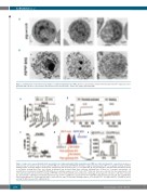
Figure 2. Transmission electron microscopy images of cultured megakaryocytes (MK). (A) Three representative annexin V microbead-isolated PSLow larger size, lower granularity MK. (B) Three representative annexin V microbead-isolated PSHigh smaller size, higher granularity MK.
A
B
CDE
Figure 3. Larger size, lower granularity (LLG) and smaller size, higher granularity (SHG) megakaryocytes (MK) are viable and apoptotic, respectively. Assays per- formed on day 13 cultures for (A-C). (A) LLG and SHG MK were stimulated with PAR4-AP, thrombin, collagen related peptide (CRP) or no agonist (resting), and PAC1 binding (marker of integrin αIIbβ3 activation) was quantified by flow cytometry (n=4). n.s.: not significant. (B) LLG and SHG were stimulated with thrombin (red line) or no agonist (resting; black line) and calcium mobilization was measured (gray lines represent Standard Error of Mean for 3 separate experiments). (C) Quantification of proplatelet forming (PPF) MK blinded as to whether samples were PSLow LLG or PSHigh SHG cells. Data were collected from five independent cords and each data point represents the percentage of PPF MK counted from randomly selected objective field image. An average 200 cells were counted per cord sam- ple. (D) Schematic outlining for DiO labeling experiment. Day 13 PSLow LLG MK were separated from PSHigh SHG MKs using annexin V microbeads. PSLow LLG MK were labeled with Vybrant DiO cell tracking dye and re-cultured for two days. (E) Bar graph showing numbers of DiO-positive PSLow LLG MKs and PSHigh SHG MK at day 13 and day 15 (48 hours post DiO addition) (n=6).
A
B
2078
haematologica | 2019; 104(10)
