
Page 181 - 2019_10 resto del Mondo_web
P. 181
BCL2L2 promotes MK numbers and PPF
V microbeads were used to separate day 13 PSLow LLG MK from PSHigh SHG MK (purity shown in Online Supplementary Figure S3). Transmission electron microscopy revealed that PSLow LLG MK exhibited many typical features of BM MK, including large cells with multi-lobed nuclei, mito- chondria, granules and surface protrusions (Figure 2A). In contrast, most of the PSHigh SHG MK were apoptotic, dis- playing membrane blebbing, highly condensed or absent nuclei, and few or no granules (Figure 2B). CD41a+ LLG MK showed a substantial increase in mitochondrial mem- brane potential compared to CD41a+ SHG MK (Figure 1D), further supporting viability of the former.
Several approaches to characterize the functionality of the LLG and SHG MK were undertaken. The signaling capacity of these different MK populations was assessed and LLG, but not SHG, were able to activate integrin αIIbβ3 in response to agonist stimulation (Figure 3A). In addition, thrombin-induced Ca2+ mobilization was observed only in LLG cells but not in SHG cells (Figure 3B). Importantly, by day 13 we observed MK proplatelet formation (PPF). After separating LLG from SHG based on PS exposure, we observed more PPF in PSLow LLG MK than in PSHigh SHG MK (Figure 3C).
Next, we sought to address whether the LLG popula- tion was the origin of the SHG cells. Day 13 PSLow LLG MK
were isolated and labeled with lipophilic cell tracking dye DiO18,19 and cultured for an additional 48 hours (h) to fol- low the 'movement' of the DiO label to SHG MK (Figure 3D). We observed an increase in DiO+ LLG MK at day 15 (Figure 3E). More importantly for this experiment, signif- icantly more SHG MK contained tracking dye after an additional two days in culture (Figure 3E). These data indi- cate that CB-derived MK cultures represent a continuum of cells transitioning from mature viable CD41aHigh CD42aHigh PSLow functional LLG MKs into CD41aLow CD42aLow PSHigh apoptotic SHG MK.
BCL2L2 regulates megakaryocyte apoptosis and larger, lower granular megakaryocyte number
To begin to understand the molecular mechanisms reg- ulating cultured MK apoptosis, we screened 24 genes with established roles in intrinsic and extrinsic apoptosis path- ways whose expression changed as MK matured in cul- ture. Because the percentages of SHG MK substantially increased between day 6 and day 13 (Online Supplementary Figure S4), we performed gene expression profiling on CD61-purified MK at these time points and observed a 3-fold and 12-fold increase, respectively, in BCL2L1 and BCL2L2, two anti-apoptotic Bcl-2 family members (Figure 4A). BCL2L1 encodes Bcl-xL and has an established role
AB
CD
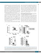
Figure 1. Two distinct populations emerge during cultured megakaryocytopoiesis. (A) Forward scatter (FSC) and side scatter (SSC) flow cytometric analysis of day 13 cultures showing the distinct larger size, lower granularity (LLG) and smaller size, higher granularity (SHG) populations of megakaryocytes (MK). (B) LLG and SHG cells were quantified for dual MK markers CD41aCD42a (n=10) and CD41aCD42b (n=10) by flow cytometry. (C) LLG and SHG MK were analyzed by flow cytometry using PE-labeled annexin V and APC-labeled anti-CD41a (n=5). (D) LLG and SHG MK were stained with APC-labeled anti-CD41a and TMRM to detect mitochondrial membrane potential and analyzed by flow cytometry (n=4). Error bars indicate mean±SEM. All assays performed on day 13 cultures.
haematologica | 2019; 104(10)
2077
