
Page 118 - 2019_10 resto del Mondo_web
P. 118
M. Chapellier et al.
ABCD
EFG
HIJ
KL
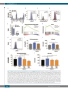
Figure 7. TNFSF13 promotes human acute myeloid leukemia cells in an NF-kB-dependent manner. (A) Output cell number of human myeloid leukemia cell lines cultured under serum-free conditions for three days with or without (Control) TNFSF13. A total of 1,000 cells were seeded per well (n=3). (B) Representative flow cytometric analysis showing staining of TNFSF13 on the cell surface of Mono-Mac-6 following 5-minute (min) stimulation with TNFSF13 (blue, 100 ng/mL) compared to non-stimulated cells (Control, white). Isotype control is shown in light gray. (C) Representative flow cytometric analysis showing SYNDECAN-1 (CD138) (dark gray) expression on Mono-Mac-6 cells. Isotype control is shown in light gray. (D) Representative flow cytometric analysis showing staining of TNFSF13 on the cell surface of Mono-Mac-6 following 5 min stimulation with TNFSF13 (100 ng/mL); with (red) or without (blue) heparin (4 IU/mL). Non-stimulated cells were used as control (white). (E) Percentage of early (Annexin-V+7AAD–) and late (Annexin-V+7AAD+) apoptotic Mono-Mac-6 cells following three days of stimulation with TNFSF13 (n=3). (F and G) Mono-Mac-6 cells were stimulated with TNFSF13 for 24 hours (h) prior to RNA sequencing. Gene Set Enrichment Analysis identified an enriched (F) TNF recep- tor signature (gene set from MsigDB, molecular signatures database) and an (G) NF-κB signature.50 (H) Phospho-flow cytometric analysis of Mono-Mac-6 cells stimu- lated with TNFSF13 for 1 h. Isotype (light gray) and pNF-kB (Control, dark gray; TNFSF13, blue) staining. (I and J) Mono-Mac-6 cells were stimulated with TNFSF13 in the presence of a TNFRSF17 blocking antibody or corresponding isotype control, and analyzed for (I) pNF-kB expression after subtracting the signal using matching isotype control antibodies, and (J) output cell number after three days. (K and L) Mono-Mac-6 cells were treated with the IKK inhibitor TPCA1 at 1 or 3 μM during TNFSF13 stimulation and analyzed after three days for (K) pNF-kB expression after subtracting the signal using matching isotype control antibodies and (L) output cell number. Values are means±Standard Deviation. *P<0.05; **P<0.01; ****P<0.0001. FDR: false discovery rate; DMSO: dimethyl sulfoxide.
2014
haematologica | 2019; 104(10)
