
Page 114 - 2019_10 resto del Mondo_web
P. 114
M. Chapellier et al.
screen, IL9 also supported the growth of leukemic cells in vitro (Online Supplementary Figure S3A) and ex vivo stimula- tion of c-Kit+ leukemia cells with IL9 prior to transplanta- tion into mice resulted in elevated levels of leukemia cells in the blood and reduced survival compared to controls (Online Supplementary Figure S3B and C). Taken together, these observations validate our barcoded screening strate- gy and demonstrate that primarily TNFSF13, but also IL9, support AML-initiating cells.
Myeloid cells secrete TNFSF13
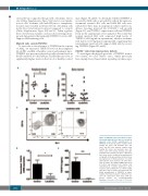
To assess the in vivo relevance of TNFSF13 in the context of AML, we measured TNFSF13 levels in the peripheral blood (PB) and BM of healthy control and leukemic mice. TNFSF13 was present at physiologically relevant levels in the PB and BM of both leukemic and healthy mice but at significantly higher levels in the blood of healthy control
AB
mice (Figure 3A and B). To determine whether TNFSF13 is secreted by AML cells or provided by cells in the microen- vironment, normal c-Kit+ cells and AML BM cells were cultured for three days in suspension cultures under con- ditions favoring myeloid (Gr-1+CD11b+) cell growth (Figure 3C), and TNFSF13 expression in cells and TNFSF13 levels in the supernatants were analyzed. We found that normal myeloid BM cells expressed high levels of TNFSF13 (>240 ng/mL in supernatant), whereas leukemic cells did not (<10 ng/mL in supernatant), suggesting that non-leukemic myeloid cells support AML cells by secret- ing TNFSF13 (Figure 3D and E).
Tnfsf13-/- mice have myelopoiesis defects
To investigate the physiological role of TNFSF13 in an in
vivo context, we used Tnfsf13-/- mice, which previously have mainly been characterized regarding cytokine regu-
C
DE
Figure 3. TNFSF13 is present in the bone mar- row and peripheral blood and is secreted by myeloid cells. ELISA quantification of TNFSF13 in (A) blood plasma samples from healthy (10 mice) and leukemic mice (12 mice) and (B) bone marrow samples from healthy (12 mice) and leukemic mice (13 mice). (C-E) c-Kit+ normal (control) and c-Kit+ leukemic bone marrow cells were cultured for three days under conditions favoring myeloid cell growth. (C) Representative dot plots show- ing expression of CD11b and Gr-1 on control and leukemic bone marrow cells after three days of culture. (D) Tnfsf13 mRNA expression in control and leukemic bone marrow cells determined by real-time polymerase chain reaction and normalized to a control sample after three days of culture (n=3 per group). (E) ELISA quantification of TNFSF13 in super- natants of normal or leukemic c-Kit+ bone mar- row cells cultured for three days (n=8 per
group). Values are
Deviation. *P<0.05; **P<0.01; ***P<0.001.
means±Standard
2010
haematologica | 2019; 104(10)
