
Page 98 - 2019_09-HaematologicaMondo-web
P. 98
J. Liu et al.
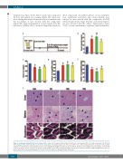
Supplementary Figure S12A). These results were supported by liver and spleen iron staining (Figure 5F). Moreover, iron staining demonstrated increased iron accumulation in the duodenum of mice treated with the compounds (Figure 5F), suggesting inhibition of iron transfer from the intestine to plasma due to increased hepcidin driven by
these compounds. As further evidence of iron redistribu- tion, transferrin saturation and serum ferritin were reduced in mice treated with the compounds (P<0.05) (Online Supplementary Figure S12B,C), and serum transfer- rin was elevated (P<0.05) (Online Supplementary Figure S12D). Serum interleukin-6, aspartate aminotransferase,
AB
CD
E
F
Figure 6. Compound administration to iron-depleted Hfe-/- mice. (A) The experimental design of treatment of iron-depleted Hfe-/- mice with compounds 93, 156 and 165. Changes in (B) serum hepcidin, (C) serum iron, (D) splenic iron and (E) hepatic iron of 9-week old Hfe-/- mice with iron depletion for 3 weeks prior to the admin- istration of compounds 93, 156 and 165 at a dose of 30 mg/kg body weight for another 2 weeks (n=4-6). (F) Tissue iron staining of liver and spleen sections with Perls Prussian blue (in blue, indicated by arrows) and duodenal sections with 3'-diaminobenzidine-enhanced Perls stain (in brown). Original magnification, ×200 for spleen, and ×400 for liver and duodenum. *P<0.05; #P<0.001, relative to untreated control (Ctrl) mice.
1776
haematologica | 2019; 104(9)
