
Page 87 - 2019_09-HaematologicaMondo-web
P. 87
A unique ABCB7-FECH-ABCB10 complex
occurred 3 days after KD of ABCB7 and accounted for the increased influx of iron in mitochondria. The decreased cytosolic aconitase activity in ABCB7-depleted cells was therefore likely the result of the conversion of IRP1 from cytosolic aconitase into IRE-binding apo-protein, rather than the result of impaired cytosolic ISC biogenesis, as pre- viously proposed.11 Similarly, yeast cells depleted of Atm1 were reported to activate the iron regulon, which encodes the high affinity iron uptake system.43,44
Despite the large amount of iron imported in mitochon- dria, cells lacking Abcb7 showed a defect in heme biosyn- thesis, which resulted not only from translational repres-
sion of Alas2 by IRP, but also from the decreased stability of ferrochelatase. Recent studies proposed that in a mouse model of frataxin deficiency, mitochondrial iron overload was mediated by the upregulation of MFRN2, which was driven by defective heme biosynthesis, due to loss of FECH.45 Interestingly, mitochondrial iron accumulation has also been reported in erythroblasts from patients with ery- thropoietic protoporphyria due to decreased ferrochelatase levels,46 suggesting that loss of FECH and/or its product, heme, in cells with an intact IRE-IRP system may drive mitochondrial iron overload.
The identification and characterization of protein–pro-
AB
C
D
E
F
G
H
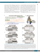
Figure 6. Mutational analysis of the C-terminal domain of ABCB7 identified the region V450-D538 as being involved in binding ferrochelatase. (A) Primary sequence of the C-terminal domain of ABCB7. (B) Modeled crystal structure of ABCB7. The numbered peptide sequences in (A) and the corresponding colored regions in (B) refer to the amino acid residues that were subjected to alanine scanning mutagenesis to assess their involvement in interacting with FECH. (C-E) Immunoprecipitation (IP) of FLAG-tagged ABCB7 wildtype (B7) or the mutants, as indicated, expressed in G1E-ER4 cells that had been silenced for 3 days to knockdown the expression of endogenous Abcb7 and that co-expressed HA-tagged FECH. Mutants 1, 2, 3, 4 and 6 of ABCB7 [green peptides and domains in (A) and (B), respectively] showed significantly decreased binding to FECH and to Abcb10. (F-H) In vitro pull-down assays of 35S-labeled FLAG-tagged ABCB7 wildtype (B7) or mutants, as indicated, in the presence of 35S–FECH (F) confirmed the results obtained in vivo (C-E) and demonstrated that binding of FECH to ABCB7 was through direct physical interaction. (C-E, n=6. F-H, n=4). See also Online Supplementary Figure S17 for densitometries of immunoblots and statistical analyses.
haematologica | 2019; 104(9)
1765
