
Page 80 - 2019_09-HaematologicaMondo-web
P. 80
N. Maio et al.
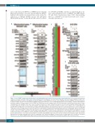
cytosol and increased MFRN1 or MFRN2 in mitochondria likely increased flux of iron into mitochondria in ABCB7- depleted cells (further investigated later in this study). Re- expression of wildtype ABCB7, but not the pathogenic XLSA E433K mutant13 in the ABCB7-KD cells restored lev-
els of FECH and SDHB, activities of complex II and aconi- tases and normalized levels of MFRN1 and IRP2 (Figure 1D and Online Supplementary Figure S2H), demonstrating that loss of ABCB7 caused profound early-onset of mito- chondrial dysfunction.
ABCD
Figure 1. Loss of ABCB7 disrupts mitochondrial function and downregulates genes involved in mitochondrial energy metabolism. (A) Protein blots and respiratory complexes I (CI) and II (CII) in-gel activity assays on mitochondrial lysates from HEK293T cells expressing an inducible control short hairpin (sh)RNA (sh_CTRL) or two shRNA targeting different regions of the ABCB7 transcript (sh_ABCB7-1 and sh_ABCB7-2) for 3 days. Levels of ABCB7, mitoferrin 2 (MTFN2), superoxide dismutase 2 (SOD2), aconitase (ACO2), CII subunits SDHA and SDHB, ferrochelatase (FECH), the CI Fe-S subunit NDUFS8, glutaredoxin 5 (GLRX5), complex IV subunit MTCO1, and citrate synthase (CS) were assayed. Levels of VDAC1 and TOM20 were used as loading controls. (B) Protein blots and CI and CII in-gel activity assays on mito- chondrial lysates from G1E-ER4 control cells or from cells depleted of Abcb7 for 3 days. Comparisons between cells at the burst-forming unit erythroid stage (undif- ferentiated, without β-estradiol) or at the orthochromatophilic stage (differentiated for 72 h with β-estradiol) are shown. Levels of Abcb7, CI Fe-S subunit Ndufs1, CII subunits Sdha and Sdhb, Fech, CIII subunit Uqcrc2, Suclg2, mitochondrial unfoldase Clpx and protease Clpp were assessed by western blot. Levels of Vdac1 and Tom20 were used as loading controls. (C) Log2-fold expression of mitochondrial energy metabolism pathway genes which were differentially expressed in G1E-ER4 cells 48 h after knockdown (KD) of Abcb7 (false discovery rate <0.01, n=3 biological replicates). (D) Complementation assays on G1E-ER4 cells silenced for 3 days to KD the expression of Abcb7 and transfected with wildtype FLAG-tagged ABCB7 or with the X-linked sideroblastic anemia pathogenic mutant ABCB7E433K-F. Levels of Abcb7, Irp2, Pold1, Fech and Mfrn1 are shown, along with CII in-gel activity assay. Tom20 and a-Tubulin (Tub) were used as loading controls for the mitochondrial fractions and total lysates, respectively. (E) Protein blots on total lysates from G1E-ER4 cells. Levels of the cytosolic and nuclear Fe-S proteins Ciapin1, Glrx3, Pold1, Dpyd and Ppat are shown, along with levels of the CIA components Ciao1, Fam96b and Mms19. Tub was used as a loading control. (A, B, D and E, n=6 biological replicates). See also Online Supplementary Figures S13 and S14 for densitometries of immunoblots and statistical analyses. NT: not treated (control).
E
1758
haematologica | 2019; 104(9)
