
Page 168 - 2019_09-HaematologicaMondo-web
P. 168
S.C. Oostindie et al.
Colocalization of cell-bound CD20 and CD37 mAbs was further examined by directly assessing molecular proximi- ty using fluorescence resonance energy transfer (FRET) analysis. We examined FRET on Daudi cells between WT and Hx variants of CD20 and CD37 mAbs alone and in combination. Consistent with its CDC activity (Figure 2A), WT IgG1-CD20-7D8 induced high FRET, which suggests antibody hexamer formation (Figure 4B). WT IgG1-CD20- 11B8 did not demonstrate proximity-induced FRET (Figure 4C), and WT IgG1-CD37 induced approximately 15% FRET (Figure 4B and C). Introducing a Hx mutation result- ed in increased FRET levels for each of the single agents, indicating that enhancing Fc-Fc interactions increases mAb colocalization at the cell surface (P<0.0001) (Figure 4D and E). Introduction of the Hx mutation did not affect target binding (data not shown), thereby excluding the possibility that increased FRET would be due to more mAb being available on the cell surface. Combinations of WT IgG1- CD20-7D8 and WT IgG1-CD37 induced approximately 30% FRET, which was increased compared to the WT IgG1-CD37 single mAb (P<0.0001, Figure 4B). Combinations of WT IgG1-CD20-11B8 and WT IgG1- CD37 substantially increased FRET compared to each sin- gle mAb (P<0.0001) (Figure 4C), consistent with the enhanced CDC induction (Figure 2B). Combinations of Hx-CD20-7D8 or Hx-CD20-11B8 with Hx-CD37 further enhanced FRET compared to the FRET levels induced by the WT mAb combinations (P<0.0001) (Figure 4D and E). These results confirm that CD20 and CD37 IgG1 mAbs bind in close proximity on the cell membrane, which can be enhanced by introducing the E430G mutation.
Hexamerization-enhanced CD20 and CD37 mAbs cooperate in complement-dependent cytotoxicity through Fc-mediated clustering in hetero-hexamers
Both enhancing Fc-Fc interactions in the CD20 or CD37 mAbs and combining the two B-cell target mAbs resulted in enhanced mAb colocalization. Together with the dependency of CDC on the formation of hexameric IgG complexes on the cell surface,15 this suggests that the CD20 and CD37 mAbs might not only form hexamers composed of mAbs bound to identical surface targets, but may coop- erate by also forming mixed hexameric complexes of mAbs bound to either target, referred to here as hetero- hexamers. The contribution of Fc-Fc interactions between Hx-CD20-11B8 and Hx-CD37 to the CDC activity of the mAb combination was examined by the introduction of the complementary Fc-Fc interface mutations K439E and S440K. K439E and S440K suppress Fc-Fc interactions between antibody molecules containing the same muta- tion, whereas Fc-Fc interactions are restored in K439K and S440K antibody mixtures.15 The capacity of Hx-CD20- 11B8 and Hx-CD37 variants with K439E and S440K muta- tions to induce CDC was tested using Daudi and WIL2-S cells. The CDC activity of Hx-CD20-11B8 was completely inhibited by introducing either the K439E or S440K Fc-Fc inhibiting mutation using Daudi and WIL2-S cells (Figure 5A and B). CDC activity was restored when Fc-Fc inhibi- tion was neutralized by mixing the two CD20 mAbs. Similar results were observed for Hx-CD37 on Daudi cells, while on WIL2-S cells, Hx-CD37 did not induce CDC, most likely due to low CD37 expression (Figure 5C and D). Combining Hx-CD20-11B8 and Hx-CD37 mAbs harbor-
AB
CD
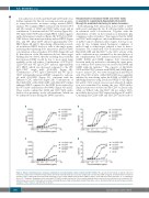
Figure 3. Enhanced binding and use of C1q by combinations of hexamerization-enhanced CD20 and CD37 mAbs. The capacity to bind C1q (A, C) and the efficiency to bind C1q and promote Complement-dependent cytotoxicity (CDC) (B and D) was assessed using Daudi cells opsonized with 10 μg/mL of hexamerization-enhanced variants of type I CD20 mAb-derived Hx-CD20-7D8 (A-B) or type II CD20 mAb-derived Hx-CD20-11B8 (C and D), CD37 mAb 37.3-derived Hx-CD37, or a combination thereof (5 + 5 μg/mL). Binding was detected using a FITC-labeled rabbit anti-human C1q secondary antibody and is expressed as mean fluorescence intensity. CDC induction was assessed in C1q-depleted serum by calculating the percentage of propidium idodide (PI)-positive cells as determined by flow cytometry. Representative examples of three replicate experiments are shown.
1846
haematologica | 2019; 104(9)
