
Page 146 - 2019_09-HaematologicaMondo-web
P. 146
B. Gonzalez-Farre et al.
to verify the presence of the 11q aberration by FISH in all ten evaluable cases (Online Supplementary Figure S3 and Supplementary Table S5). The morphological, clinical, and genetic features and consensus diagnosis of the 11 BLL- 11q identified in our files are summarized in Table 1. The six cases negative for the MYC rearrangement and 11q aberrations by Oncoscan were re-classified as DLBCL (3 cases) or HGBCL, NOS (3 cases). The DLBCL had pre- dominant centroblastic morphology, germinal center phe- notype, very high proliferative index and a focal “starry sky” pattern (see Online Supplementary Results). The absence of 11q alterations was also verified using the 11q FISH probe in four of these MYC/11q-negative cases with available material (Online Supplementary Figure S1A).
tumors were reclassified morphologically as HGBCL, NOS, two as DLBCL and only one case was considered atypical BL. None of the cases was considered as typical BL (Figure 1). Six cases showed a “starry sky” pattern and two had a nodular growth pattern with the presence of a disrupted follicular dendritic cell meshwork (Figure 1C). Ki67 was very high in all the samples, as in BL. All cases had a germinal center (GC) phenotype and GC B-cell (GCB) signature by Nanostring Lymph2Cx assay. MUM1/IRF4 was negative in all 11 cases. One case expressed BCL2 (Figure 1D). LMO2, a germinal center marker that is usually seen in GCB-DLBCL but not in BL18 was expressed in five cases (Figure 1A, B). Interestingly, using a 40% cutoff,19 five cases were positive for MYC expression. However, only one case showed diffuse and intense positivity while the other four cases had either only positivity in around 50% of the cells or the intensity was not that expected in typical BL. Additionally, MYC RNA levels were significantly lower in BLL-11q than in MYC-positive BL (relative expression 0.07 vs. 0.36, P=0.019) (Online Supplementary Figure S4A). Epstein-Barr virus hybridization was negative in the nine cases tested.
Clinically, BLL-11q frequently had a nodal localized presentation (8/11) in the head and neck region. Two cases had an extranodal presentation, one in the context of an acute appendicitis and the other debuted as an omental mass. Eight patients (73%) had stage I-II, and one patient presented in an advanced stage (IV-E) with
In the 35 older patients (≥40 years old), a MYC translo- cation was found in 32 cases; one was classified as DLBCL, 21 as BL, and ten were HGBCL with double- or triple-hit aberrations (BCL2 and/or BCL6 translocations). Only three cases were negative for MYC translocations and were classified as HGBCL, NOS (Online Supplementary Figure S1B and Online Supplementary Results). We screened these cases with the 11q FISH probe and all three were negative for the 11q aberration.
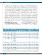
Clinical and morphological results of cases of Burkitt-like lymphoma with 11q aberration
The 11 patients with BLL-11q had a mean age of 15 years (range, 8-37 years); eight were male (Table 1). Eight
Table 1. Pathological and clinical features of 11 cases of Burkitt-like lymphoma with 11q aberration.
Case Age/ gender
#1 27,M
Biopsy site
Laterocervical LN
Axillary
LN Tonsil
Original Final diagnosis diagnosis
Immunophenotype
CD10& IRF4/ BCL2 LMO2 MYC BCL6 MUM1
Stage*
COO Chemo- Rituximab Outcome/
Atypical BL
Atypical
BL HGBCL
HGBCL
HGBCL
HGBCL
HGBCL, NOS
DLBCL
DLBCL & HGBCL blastoid
HGBCL,NOS
HGBCLwith features intermediate between BL and DLBCL
DLBCL
+ ---+I
Nanostring therapy (Lymph2Cx)
GCB A Yes
GCB A Yes
GCB P No
follow-up
CR, 72m
CR, 112m
CR, 54m
#2**
#3
#4
#5
#6 #7
#14
37,M
8,F
+ --
+ --
+ - -
+ - +
+ - -
+ - - - - I
+ - - - Weak+ II
17,F Submaxilar
+-IV-E
- - II
+ + I
+ - I
GCB A
Yes CR,22m
14,F
14,M 8,M
8,M
LN
Laterocervical LN
Appendix
+-II
GCB P No CR,29m
GCB P No CR, 25m GCB P No CR,113m
GCB P No CR, 15m
GCB P No CR,35m
GCB P Yes CR, 12m
GCB A Yes CR,4m
Laterocervical BL AtypicalBL LN
Laterocervical BL
LN
Laterocervical DLBCL mass
Laterocervical DLBCL
LN
Omentum HGBCL
HGBCL blastoid
#15 12,M
#16 14, M
#17 16,M
HGBCL,NOS +
HGBCL, NOS +
HGBCL,NOS +
- - - + I
- - +-III
- - - + III
*Stage was established according to the St. Jude/International Pediatric NHL Staging System (IPNHLSS) or Ann Arbor staging system for pediatric and adult patients, respectively. COO: cell of origin; M: male; F: female; LN: lymph node; BL: Burkitt lymphoma; HGBCL: high-grade B-cell lymphoma; NOS: not otherwise specified; DLBCL: diffuse large B-cell lymphoma; Epstein-Barr virus in situ hybridization (EBER) was negative in all nine tested cases. E: extranodal; GCB: germinal center B-cell; A: adult schema protocol (R-CHOP or burkimab); P: pediatric schema protocol; CR: complete response; m: months. All patients received central nervous system prophylaxis. **Human immunodeficiency virus (HIV)-positive patient
1824
haematologica | 2019; 104(9)
