
Page 174 - 2019_08-Haematologica-web
P. 174
X. Zhu et al.
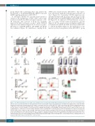
siRNA were transfected into MSC-ITP-C+. The expres- sion of IL-1β was markedly reduced in the cell lysates on day 7 (Figure 3G). Conversely, CXCL12 expression was upregulated (Figure 3H). IL-1β knockdown decreased the phosphorylation of MyD88, ERK1/2, p38 MAPK and NF- κB (Figure 3I). As expected, CCK8 proliferative assays at days 1, 3 and 7 revealed altered proliferative capacity (Figure 3J, K). The apoptosis rate was higher in MSC-con- trol transfected with IL-1β cDNA and lower in MSC-ITP- C+ with IL-1β-siRNA (Figure 3L, M). Furthermore, bone marrow sections from patients in the MSC-ITP-C+ group
ABCD
groups (Figure 3D). Collectively, these data indicate the involvement of IL-1R/MyD88/NF-kB, ERK1/2 and p38 MAPK signaling pathways in MSC-ITP-C+.
To determine whether the autocrine IL-1β protein is crucial for the dysfunction of MSC, MSC-control were transfected with IL-1β-cDNA and signal sequences of IL- 1R antagonist. The secretion of IL-1β was markedly improved in MSC cell lysates on day 7, while CXCL12 expression was inhibited (Figure 3E, F). Moreover, phos- phorylation of MyD88, ERK1/2, p38 MAPK and NF-κB was enhanced (Figure 3I). Lentiviruses carrying IL-1β-
E
F
GH
I
JL
KM
Figure 3. The C5b-9/interleukin-1β loop regulates mesenchymal stem cells from patients with immune thrombocytopenia. (A, B) Levels of pro–interleukin-1β (IL- 1β) and caspase 1 (A) and interleukin-1 receptor (IL-1R) (B) in the three groups [MSC-control: mesenchymal stem cells (MSC) from healthy subjects; MSC-ITP-C- MSC from patients with immune thrombocytopenia (ITP) without complement deposition on MSC; MSC-ITP-C+: MSC from patients with ITP with complement deposition on MSC] were detected by western blotting in MSC cell lysates. β-actin was used as the loading control (MSC-control, n=12; MSC-ITP-C-, n=12; MSC-ITP-C+, n=12; One-way analysis of variance (ANOVA). (C, D) Phosphorylation of MyD88, NF-κB, ERK1/2 and p38 MAPK signaling pathway proteins in MSC from ITP patients and healthy volunteers. β-actin was used as the loading control (MSC-control, n=12; MSC-ITP-C-, n=12; MSC-ITP-C+, n=12; one-way ANOVA). (E, F) Expression of IL-1β and CXCL12 in MSC-control cell lysates at day 1 and day 7 with transfection with lentivirus carrying the IL-1β-cDNA from two independent experiments (n=12; Student t-tests). (G, H) Expression of IL-1β and CXCL12 in MSC-ITP-C+ cell lysates 7 days after transfection with lentivirus carrying the IL-1β-siRNA from two independent exper- iments (n=12; Student t-tests). (I) Phosphorylation of MyD88, ERK1/2, p38 MAPK and NF-κB in transfected MSC by Western blotting (n=8; Student t-tests). (J, K) CCK8 proliferative assays of transfected MSC from three independent experiments (MSC-control, n=12; MSC-ITP-C+, n=12; Student t-tests). (L, M) Apoptosis rate of transfected MSC (MSC-control, n=12; MSC-ITP-C+, n=12; χ2 tests).
1666
haematologica | 2019; 104(8)
