
Page 92 - 2019_07 resto del Mondo-web
P. 92
R.G. Morgan et al.
HEL cell lines. This approach resulted in a 72%±7% knockdown in LEF-1 protein in the nuclei of K562 cells and an 89%±4% knockdown in HEL cells (Figure 6A). Somewhat lower levels of LEF-1 knockdown were observed in CHIR99021-treated cells (65%±19% and 83%±7%, respectively) probably a result of LEF1 being a Wnt target gene and thus being induced through Wnt ago- nist treatment.39 LEF-1 knockdown perturbed nuclear localization of β-catenin by approximately one-third (28%) in K562 following CHIR99021 treatment, propor- tionate to control cells. This reduction was accentuated in HEL cells (41%) which corresponded to the greater degree of LEF-1 knockdown in these cells (Figure 6B). The knock- down of LEF-1 protein resulted in significantly reduced growth of both K562 and HEL cells at multiple time points across a range of serum concentrations (Figure 6C). Use of a second LEF1 shRNA and a different method of Wnt stimulation (rWnt3a) resulted in a similar finding (Online Supplementary Figure S7B and C). These data suggest LEF-1 promotes the optimal translocation of β-catenin into the nucleus of Wnt-responsive cells and partly contributes to their growth.
Next, we examined whether LEF-1 expression was suf- ficient to permit nuclear-localization of β-catenin. To
AB
establish this, we stably over-expressed LEF1 in the Wnt- unresponsive (and LEF-1 negative) U937 and ML1 cells. Overexpression of LEF-1 resulted in substantial cytosolic expression of the full-length LEF-1 protein (50kDa) but weak nuclear expression; despite this, we observed a dra- matic increase in nuclear localized β-catenin in both ML1 (4-fold) and U937 (2.3-fold) cells over-expressing LEF1 fol- lowing CHIR99021 treatment (Figure 6D and E). This dis- parity may be explained by the abundant expression of a short-form of LEF-1 in the nucleus (25-30kDa) that was absent in Wnt-responsive lines (discussed below). These effects were mirrored using Wnt3a treatment (Online Supplementary Figure S7C) and we also showed that LEF-1 overexpression was able to facilitate nuclear localization of β-catenin in two further AML cell lines (PLB-985 and THP1) (Online Supplementary Figure S7D). These data demonstrate that overexpression of LEF-1 can significantly increase the capacity for nuclear β-catenin localization in Wnt-unresponsive cell lines.
To assess the impact of LEF-1 knockdown (and subse- quent nuclear β-catenin reduction) on Wnt signaling, we measured TCF reporter activity. As predicted, Wnt signal- ing induction in Wnt-responsive K562 and HEL cells fol- lowing CHIR99021 treatment was severely diminished
CD
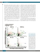
Figure 3. Proteomics analyses reveal contrasting β-catenin inter- action profiles between Wnt- responsive and Wnt-unresponsive leukemia cell lines. Scatter plots showing β-catenin protein interac- tions detected in (A) K562 cytoso- lic, (B) K562 nuclear, (C) ML1 cytosolic, and (D) ML1 nuclear frac- tions. Vertical dashed red line indi- cates the threshold for 2-fold change in protein binding at log2 (=1) relative to IgG co-immunopre- cipitation.. Horizontal red line rep- resents threshold for significant interactions at P=0.05 on log10 scale (=1.3). Highlighted red dots indicate statistically significant interactions and green highlighted events/labels indicate known inter- actions/associations for β-catenin. Remaining black dots represent other proteins detected in the MS
analysis; see
Supplementary Data sheets. Fold change values less than 0 are not shown because these likely repre- sent contaminants (see Online Supplementary MS data sheets).
also Online
1370
haematologica | 2019; 104(7)
