
Page 89 - 2019_07 resto del Mondo-web
P. 89
LEF-1 regulates β-catenin nuclear localization
v.10.5.3 (Tree Star Inc., Ashland, OR, USA). Threshold for TCF reporter fluorescence was set using matched-controls expressing mutant ‘found unresponsive’ fuBAR. Cell viability was assessed using 2 μg/mL propidium iodide (Miltenyi Biotech).
Statistical analysis
Statistical analyses were performed using GraphPad Prism v.7.0 (GraphPad Software Inc., San Diego, CA, USA) and Perseus. Correlation was assessed using a Spearman Rank correlation coef- ficient (R). Significance of difference was assessed using a one- sample or Student t-test and data represents mean±one Standard Deviation (SD) derived from three biological replicates.
Results
Myeloid leukemia cell lines exhibit heterogeneous nuclear β-catenin localization and Wnt activation
Previously we showed that myeloid leukemia cell lines vary markedly in their capacity for nuclear β-catenin local-
A
B
CD
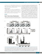
ization upon Wnt stimulation, mimicking the heterogene- ity of nuclear β-catenin translocation in AML patients.11 To investigate the mechanistic basis for this, we selected two sets of myeloid leukemia lines which differed markedly in Wnt signaling output in response to agonist. K562 and HEL were Wnt-responsive cell lines that localized high levels of β-catenin into the nucleus upon treatment with the Wnt agonist, CHIR99021, a GSK3β inhibitor (Figure 1A). Cell viability was not significantly affected by 16 h CHIR99021 treatment (Online Supplementary Figure S1A and B). Similar patterns of subcellular localization were observed for the active (non-phosphorylated) form of β- catenin in response to CHIR99021 (Online Supplementary Figure S1C). As expected, the phosphorylated forms of β- catenin (Ser33/37/Thr41) were reduced upon CHIR99021 treatment (Online Supplementary Figure S1C). Correspondingly, these lines showed robust induction of a TCF reporter (a measure of β-catenin-dependent transcrip- tion), whilst cells expressing reporter with a mutated TCF
Figure 1. Myeloid leukemia cell lines exhibit a heterogeneous response to Wnt stimulation. (A) Representative immunoblots showing total β-catenin subcellular- localization in myeloid cells following CHIR99021 treatment (GSK3β inhibitor). Lamin A/C and a-tubulin indicate the purity/loading of the nuclear (N) and cytosol (C) fractions, respectively. (B) Representative flow cytometric histograms showing intensity of the TCF-dependent expression of YFP from the ‘β-catenin activated reporter’ (BAR) reporter, or negative control ‘found unresponsive β-catenin activated reporter’ (fuBAR) control (containing mutated promoter binding sites) following treatment with CHIR99021/vehicle control [dimethyl sulfoxide (DMSO)] as above. (C) Summary showing the relative percentage nuclear β-catenin localization (as a proportion of the total) induced in myeloid cell lines upon CHIR99021 treatment. (D) Summary showing the median fluorescence intensity generated from the BAR/fuBAR reporters in myeloid cell lines treated ± CHIR99021. *P<0.05; ****P<0.0001; ns: not significant. MFI: mean fluorescence intensity.
haematologica | 2019; 104(7)
1367
