
Page 213 - 2019_07 resto del Mondo-web
P. 213
Gradient-dependent inhibition of GPCR
AB
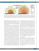
Figure 7. Proposed role of gradient-dependent inhibition in creating and maintaining the core-shell thrombus architecture. (A) As gradient-dependent inhibition (GDI) potently inhibits thrombin-induced platelet activation over a narrow range of temporal concentration gradients, we propose that stimulatory signaling via the PAR receptors will effectively be shut off outside the thrombus core. (B) In contrast, as GDI exhibits a gradual increase over a larger range of gradients for ADP and thromboxane A2, paracrine signaling from these agonists will only be partially inhibited, resulting in an intermediary state of platelet activation in the thrombus shell.
cAMP-dependent pathway, as ADP is known to decrease cAMP levels via inhibition of adenylyl cyclase.30 Lastly, GDI-platelets exhibited altered levels of total serine phos- phorylation and altered cytoskeletal organization in com- parison with resting and activated platelets, indicating that GDI involves the activation of unique kinase-dependent signaling pathways.
Our results may, at least in part, explain previous find- ings of non-responsive, “exhausted”, platelets in different situations characterized by excessive diffusion of soluble platelet agonists, for example in cancer,31-34 sepsis,34 and intensive care.35-37 However, the term “exhausted” does not fit with the findings in this study, as GDI-platelets retain the capacity to be activated by other agonists or by higher agonist gradients. The concept of GDI could also help to explain recent findings from in vivo experiments, showing the presence of a remarkably stable thrombus architecture encompassing a large shell of loosely attached, P-selectin-negative platelets with little calcium mobilization surrounding a highly activated cluster of platelets in the core of the thrombus.4,10 GDI could be one mechanism responsible for maintaining low-grade acti- vation in platelets forming the thrombus shell, despite the inevitable slow leakage of ADP, thromboxane A2 and thrombin,38 eventually leading to agonist accumulation outside the core (Figure 7A,B). In this context, it is inter- esting to note the different effects of GDI on paracrine signaling from ADP and thromboxane on the one hand and signaling from the thrombin receptors PAR1 and PAR4 on the other. Whereas GDI had incremental inhibitory effects on paracrine stimulation over a large range of gradients, resulting in progressively weaker platelet activation, a bimodal effect distribution was observed for PAR-mediated signaling, as platelet activa- tion was effectively shut off when gradients decreased below a certain threshold. These differential effects of GDI could be instrumental for the formation of the core- shell thrombus architecture, as GDI could result in abol-
ished thrombin signaling outside the thrombus core, whereas the gradual effects of GDI on paracrine stimula- tion would result in the intermediary platelet activation state found in the thrombus shell. The observation that the collagen activation pathway (represented by CRP- XL) was unaffected by GDI in our study is noteworthy in this context, as collagen is not a diffusible agonist but remains attached to the damaged vessel wall upon injury. Thus, blood exposure to collagen is inherently restricted to the immediate vicinity of vessel damage, rendering GDI physiologically irrelevant as a regulatory mecha- nism.
While this study focused exclusively on the gradient- dependent effects of single agonists on platelet activation, platelets circulating near a vessel injury are exposed to multiple stimulatory gradients, primarily including the agonists ADP, thromboxane A2 and thrombin. Adding another layer of complexity, these pro-hemostatic signal- ing pathways are counter-balanced by inhibitory signals released from intact endothelium such as PGI2 and nitric oxide. Thrombin receptors are quite common in the human body, especially on cells in circulation. The pres- ence of GDI in epithelial cells suggests that this phenom- enon may be exhibited by other cell types.
Acknowledgments
We are grateful to all the students for performing minor parts of the experiments in this study during their dissertations in our laboratory. The authors wish to thank Kjersti Claesson, a Ph.D. student in our group, for helping with microscopy and Maria Wallstedt, a research engineer, for her help with flow cytometry. We are grateful to Magnus Grenegård, Professor, at Örebro University for sharing reagents, interesting discussions and valuable suggestions during the study. We thank the Swedish Heart-Lung Foundation (grant n. 207-0440), the Swedish Research Council (grant n. 2017-01177) and the British Heart Foundation (grant n. RG/15/2/31224 to JMG) for research funding.
haematologica | 2019; 104(7)
1491
