
Page 209 - 2019_07 resto del Mondo-web
P. 209
Gradient-dependent inhibition of GPCR
flow cytometry. Receptor density was found to be unchanged in GDI-platelets, indicating that receptor inter- nalization is not a major feature of GDI (Online Supplementary Figure S9). A role for clathrin-mediated receptor endocytosis was also excluded, as GDI was unaf- fected by pre-treatment with the dynamin inhibitor Dynasore21 (Online Supplementary Figure S10). Western blotting revealed a prominent phosphorylation of VASP at serine 157 in GDI-platelets, which was not observed in resting or activated platelets (Figure 4A,B). As PKA is involved in mediating phosphorylation at S157,22 we examined the effects of PKA inhibition on VASP phospho- rylation and GDI as measured by aggregometry. Inhibition of PKA by 30 μM H89, which inhibited VASP phosphorylation by 10 nM but not 100 nM prostacyclin (PGI2) (Online Supplementary Figure S3B), did not in itself cause aggregation of platelets, nor did it affect aggregation induced by rapid (2 s) addition of 30 μM PAR1-AP. However, the effects of GDI on PAR1-induced platelet aggregation were partially reversed (Figure 4C). This effect was also reflected in a decreased level of VASP phospho- rylation in PKA-inhibited GDI-platelets treated with PAR1-AP (Figure 4A,B). Furthermore, western blotting revealed markedly decreased AKT phosphorylation in GDI-platelets in comparison with that in activated platelets (Figure 4D).
As both PKA and VASP are components of the cAMP/ adenylyl cyclase pathway, we examined the roles of adenylyl cyclase and cAMP in GDI. Pre-treatment of platelets with low doses of PGI2 (concentrations 0.01, 0.1 and 1 nM) to increase adenylyl cyclase activity did not
affect aggregation induced by a 2 s infusion of 30 μM PAR1-AP, but significantly and dose-dependently enhanced GDI at the 80 s and 160 s infusion times (Figure 4E). Similarly, pre-incubation of platelets with the phos- phodiesterase-3 inhibitor milrinone (3 μM) to inhibit cAMP degradation had no effect on aggregation at the 2s infusion time, and did not affect platelet aggregation induced by CRP-XL, at either the 2 s or the 1,280 s infu- sion time (Figure 4F). In contrast, significant potentiation of GDI was observed for PAR1-AP, PAR4-AP and ADP, with a similar trend for U46619, although the effect did not reach significance using this agonist. Treatment with epinephrine (0.1, 1 and 10 μM) to inhibit adenylyl cyclase 60 s before starting agonist infusion with PAR1-AP or thrombin did not in itself cause any aggregation, but pro- duced a significant dose-dependent inhibition of GDI (Figure 4G,H). This effect was most prominent for throm- bin, as 1 μM epinephrine was sufficient to block GDI completely for all tested infusion times.
The role of the VASP/PKA pathway in cytoskeleton remodeling has been described previously.23 Also, VASP has been shown to interact with F-actin and regulation of F-actin rearrangement is modulated by differential phos- phorylation of VASP.24 we, therefore, assessed morpholog- ical and cytoskeletal changes in platelets induced by GDI using fluorescence microscopy with staining for the cytoskeletal protein F-actin. Compared to resting and acti- vated platelets, GDI-platelets displayed a preferential dis- tribution of F-actin filaments near the cell membrane (Figure 5A). To confirm this finding and obtain more insights into the structural characteristics unique to GDI-
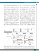
A
B
C
Figure 3. Agonist-specific effects of gradient-dependent inhibition. (A) Algorithm for defining Cres, the minimal concentration required to induce aggregation in platelets showing gradient-dependent inhibition (GDI). (B) GDI was induced by exposing platelets to Cagg with ΔCnres for the agonists ADP, PAR1-AP and PAR4-AP. Platelets were then challenged with the same agonist by adding multiples of Cagg with an infusion time of 2 s. (C) To investigate whether the determinant of the aggre- gation induced by adding PAR1-AP or PAR4-AP at the concentration Cres, as shown above in (B), was the increased agonist gradient or the final agonist concentration, subsequent infusions of Cres, using either the 2 s high gradient (1) or the GDI gradient ΔCnres (2), were performed.
haematologica | 2019; 104(7)
1487
