
Page 146 - 2019_07 resto del Mondo-web
P. 146
A. Uchida et al.
non-DH DPL and other GCB-like DLBCL cases not only indicates the abundant expression, but also implies the presence of distinct functions from its anti-apoptotic activ- ity in these groups.28,35 Furthermore, our in vitro study con- firmed that the inhibition of MCL1 by S63845 did not have any additive pro-apoptotic effect to that of veneto- clax in the two DH-DPL cell lines, whereas S63845 restored the apoptotic sensitivity to low concentrations of venetoclax in the non-DH DPL-cell line BJAB. A previous study showed that overexpression of MCL1 is observed more frequently in ABC-like DLBCL than in GCB-like DLBCL.36 Our observations indicate that the anti-apoptot- ic activity of MCL1 for DH-DPL seems to be less limited than that for any other type of GCB-like DLBCL. DH-DPL seems to be dependent mainly on BCL2 for survival, and the pro-apoptotic action of venetoclax in DH-DPL cells may be less affected by intrinsic MCL1 expression.
The BRD4 inhibitor JQ-1 is considered a promising drug for the treatment of MYC-driven lymphomas.16,17 Although JQ-1 suppressed cell proliferation, this agent did not lead to adequate apoptosis in the two DH-DPL-cell lines. Since BET inhibitors can induce cell-cycle arrest,37 this raises concerns that JQ-1 might fail to exterminate malignant cells. Although the combination with BH3
AB
mimetics is supposed to be a promising strategy, antago- nistic effects have been observed.15 BET inhibitors are effective at inhibiting tumor expansion, but might be inap- propriate for eradicating of residual lymphoma cells.
The antitumor effect of venetoclax has mainly been dis- cussed with regard to its physiological association with BCL2.21,22,24 In the present study, we demonstrated for the first time that venetoclax not only disrupts the association between BCL2 and BIM, but also modulates signal trans- duction related to BCL2 and MCL1 in DH-DPL cells. Phosphorylation at Ser70 and its mutant in the BCL2 loop domain has been shown to enhance binding to BIM and BAK, and confirmed to inhibit the effects of BH3 mimetics on its replacement of BIM in leukemia cells.27 Although protein levels of phosphorylated BCL2 are relatively high, venetoclax clearly led to dephosphorylation of BCL2 and effectively induced apoptosis in Karpas231 and OCI-Ly8 cells. In both DH-DPL cell lines, the dephosphorylation was probably mediated through accumulation of PP2A B56a in BCL2. The increased binding of PP2A B56a to BCL2 is likely to promote dephosphorylation of this pro- tein. Consequently, a considerable part of phosphoryla- tion at Ser70 seemed to be removed within 6 h after expo- sure to venetoclax. Dephosphorylation of BCL2 should
CD
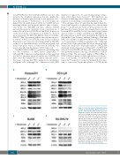
Figure 5. Venetoclax leads to dephosphorylation of BCL2 and downregulates MCL1 expression in the two double-hit and double-protein-expression lymphoma cell lines. (A-D) Although BCL2 protein levels were unchanged at least 6 h after exposure to venetoclax in Karpas231 (A) and OCI-Ly8 (B) cells, the proportion of phosphorylated BCL2 (pBCL2) was clearly decreased, and expression levels of BIM were also decreased 3 h after expo- sure to venetoclax in both double-hit and double- protein-expression lymphoma (DH-DPL) cell lines (A, B). Although the BCL2 protein levels were slightly decreased, the proportion of pBCL2 was increased 6 h after to venetoclax in BJAB cells (C). Unexpectedly, MCL1 showed decreased protein expression within 3 h after exposure to venetoclax in Karpas231 (A) and OCI-Ly8 (B) cells, but not in BJAB (C) and SU-DHL10 (D) cells. PP2A B56a showed decreased expression within 3 h after exposure to venetoclax in both DH-DPL cell lines (A, B), but not in BJAB (C) and SU-DHL10 (D) cells. In contrast, protein expression levels of PP2A B56δ were unchanged in all cell lines (A-D).
1424
haematologica | 2019; 104(7)
