
Page 115 - 2019_07 resto del Mondo-web
P. 115
SP enriches for LPC and Wnt expression in zebrafish ALL
difficult to overcome. This is largely due to the simple fact that CSC are not equivalent to normal stem cells, and as such many of the markers used to isolate stem cells in spe- cific tissues fail or underperform at isolating CSC. There is evidence that those tissue stem cell markers that do effi- ciently enrich for CSC in specific cancers, such as glioblas- toma42 and acute myelogenous leukemia,43,44 are derived from transformed stem or early progenitor cells, although this point is still controversial. In malignancies without established CSC markers, such as ALL, the CSC can only be identified retrospectively, after it has demonstrated its ability to regrow a heterogeneous tumor, at which point the original cell is lost within the bulk tumor. This study establishes that the SP assay is a promising method for enriching LPC from ALL tumors, bypassing the lack of a reliable surface marker for isolation. This assay yields only an enrichment: not every LPC is found in the SP, nor are the SP cells exclusively LPC. However, this study demonstrates that the SP can be studied as a proxy for LSC to better understand their molecular drivers and, eventually, to identify better markers.
The Rag2-Myc zebrafish model has been extensively
AB
studied in the context of T-ALL, and because the tumors derive from the thymus, it was thought to produce ALL exclusively of T-cell origin. However, a recent study from Langenau’s laboratory has provided evidence for the exis- tence of tumors of B-cell origin produced with this method, in addition to T-cell tumors.45 The origin of these B-ALL remains unclear, as they could be derived from early lymphoid progenitors located in the thymus, extra- thymic B-cell progenitors that home to the thymus after transformation, or possibly from B-cell precursors that line the thymus at later stages of development.46 Regardless, these tumors present a new opportunity to study B-ALL in zebrafish, a tumor type with few animal models.47 Another closely related model utilizing the human MYC oncogene was also recently demonstrated by Frazer’s team to produce both pre-B-ALL and T-ALL.48 Analysis of our own RNA-sequencing data showed a clear difference in expression of putative B- and T-cell genes between individual tumors, suggesting that ALL of both B- and T-cell origin are included in our dataset (Online Supplementary Figure S1). Our data indicate that SP cells from both T- and B-ALL share a common genetic sig-
C
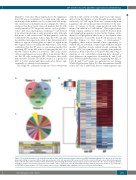
Figure 4. Sorted side population cells from different tumors share similar expression profiles and increased Wnt signaling pathway gene expression. (A) Venn dia- gram representing the overlap of differentially expressed genes in the side population (SP) versus non-SP for three tumors: 272 genes were differentially regulated between the SP and non-SP in all three tumors. (B) Heatmap demonstrating differentially expressed genes between SP and non-SP cells. Cluster analysis performed using gene analysis set from mean expression across all three tumors (761 genes, listed in Online Supplementary Table S5). Expression shown as log2 fold change. (C) PANTHER pathway analysis of 272 differentially expressed genes highlighting Wnt signaling as the most overrepresented pathway.
haematologica | 2019; 104(7)
1393
