
Page 91 - 2019_04-Haematologica-web
P. 91
New Drosophila model for chronic myeloid leukemia
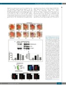
high levels of expression in some but not all photorecep- tors,14 producing a mild rough eye similar to the one observed by Fogerty7 (Figure 1A-E). This suggests that BCR-ABL1 interferes with eye development as described for the human/fly chimera. To drive BCR-ABL1 expres- sion in more eye cells, we used the glass multimer reporterGal4 (gmrGal4) driver, active in all cells committed to differentiation and located posteriorly to the morpho- genetic furrow,15 the cell indentation crossing the eye pri-
mordium from posterior to anterior (Figure 1N,O). BCR- ABL1 expression in these cells produced a severe “glazed” eye phenotype (Figure 1F-J, Online Supplementary Figure S1A,B,H,I). The regular structure of the eye was almost completely lost: ommatidia, the functional units of the eye, failed to differentiate and were no longer distinguishable. The eye was smaller, bar-shaped and misplaced extra sensory bristles appeared in the dorsal region (Figure 1H-J). Western blot
ABCDE
FGHIJ
K
LM
Figure 1. BCR-ABL1 expression in the devel- oping eye cells affects photoreceptor differ- entiation. (A-E) Adult eyes expressing EGFP (A) or BCR-ABL1 in four independent trans- genic lines, 1M (B), 3M (C), 4M (D), or 7M (E), in a subset of differentiating photore- ceptor cells under the control of the sevenlessGal4 driver construct. (C-E) High levels of BCR-ABL1 induce a “rough” eye phenotype due to impairment of cell differ- entiation. (F-J) Adult eyes expressing EGFP (F) or BCR-ABL1 (G-J) in all differentiating eye cells under the control of the gmrGal4 driver construct. (H-J) BCR-ABL1 expressed at high level in all differentiating eye cells profoundly disrupts ommatidia development inducing a “glazed” phenotype, depigment- ed area and the appearance of extra bristles (black arrows). (K-M) Quantification of BCR- ABL1 expression (K,L) and tyrosine-phos- phorylation (K,M) in protein extracts from adult heads of flies expressing either EGFP (lane 1) or BCR-ABL1 in independent trans- genic fly lines (lanes 2-5). The protein extracts were probed with antibodies raised against BCR, phosphorylated tyrosine residues (p-Tyr) or α–tubulin as the loading control. (N) Schematic of the eye-antenna imaginal disc from a late third instar larva; the positions of the eye and antenna primor- dia and of the morphogenetic furrows are indicated. The eye imaginal disc area poste- rior to the morphogenetic furrow, made of cells committed to terminal differentiation, is indicated in green. The thin black square indicates the region of interest shown in panels O-T. (O,P) Eye imaginal disc from wild- type late third instar larvae expressing EGFP under the control of the gmrGal4 driver in cells posterior to the morphogenetic furrow and expressing the pan-neuronal marker Elav in cells committed to terminal differen- tiation. (Q-T) Elav expression in eye imaginal discs from third instar larvae of the four independent transgenic lines that express BCR-ABL1 under the control of the gmrGal4 driver construct. BCR-ABL1 expression reduces the number of differentiated pho- toreceptors as indicated by the decrease of Elav-expressing cells.
N
OP
QRST
haematologica | 2019; 104(4)
719
