
Page 43 - 2019_04-Haematologica-web
P. 43
PGE2, EGF, FLT3L and TPO in human hematopoiesis
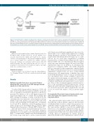
Figure 1. Xenograft model for evaluation of radioprotective substances. Cord blood-derived human CD34+ cells were transplanted into sublethally irradiated 5-week old Rag2−/−γc−/− mice. Four weeks later, xenograft mice were again irradiated with 3 Gy in order to subject human hematopoiesis to sublethal stress. Subsequently, mice were treated intraperitoneally (i.p.) once daily with the indicated molecules. Control mice were treated with the respective carrier solution (saline or ethanol). At day 8 after second irradiation, mice were sacrificed for analysis. Single cell suspensions were obtained from bone marrow and spleen. h: hours; hu EGF: human epi- dermal growth factor; mu EGF: murine epidermal growth factor; hu dmPGE2: human 16,16-dimethyl-PGE2; hu FLT3L: human FLT3L; TPO: human thrombopoietin.
RT-MLPA
RNA was isolated with Fast-Spin columns (ZymoResearch). For RT-MLPA (MRC Holland, R011-C1), specific mRNAs were reversely transcribed into cDNA and bound by two oligonu- cleotides consequently ligated. The generated amplification prod- ucts of unique length were separated by capillary sequencer (Genescan). Analysis was performed with Sequence Pilot (JSI Medical Systems). The sum of all peak data was set to 100% to normalize for fluctuations between different samples, and single peaks were calculated relative to 100%.
Statistical analysis
Statistical analysis was performed using the Mann-Whitney- Test (Graphpad Prism). P<0.05 was considered statistically signif- icant.
Results
Epidermal growth factor does not protect human hematopoietic stem cells from DNA damage-induced apoptosis in vitro and in vivo
To induce DNA damage-induced apoptosis, CD34+ cells were treated with the topoisomerase inhibitor etoposide (0.5 or 1 mg/mL) for 24 and 48 h. Apoptosis could not be prevented by addition of 20 or 200 ng/mL human EGF, respectively (Figure 2A). To exclude the possibility that EGF contained in medium or serum or produced by the cells themselves was sufficient to reduce apoptotic suscep- tibility even in the absence of accessory EGF, cells were treated with neutralizing EGF-R-antibodies (cetuximab) for 24 and 48 h. Inhibition of EGF-R-signaling did not induce apoptosis by itself (Figure 2B) nor increase DNA damage-induced apoptosis of human HSPCs (Figure 2C).
Since the effects of EGF on murine hematopoiesis were exclusively shown in vivo,10 we performed analogous experiments in a xenograft model using immunodeficient Rag2-/-γ-/- recipient mice. Animals were xenotransplanted at five weeks of age with 3x10⁵ cord blood-derived CD34+
cells. Upon successful human engraftment, mice were irra- diated with 3 Gy to mimic myelosuppression occurring during therapeutic irradiation. Subsequently, mice were treated daily with human EGF (0.5 mg/g, i.p.) or saline (100 ml, i.p.) for seven days. Regeneration of human hematopoiesis, as defined by percentage and cell count of human CD45+ cells, was determined in BM and spleen seven days after irradiation (Figure 2D and E and Online Supplementary Figure S1). In addition, we determined the proportion of immature CD34+ cells within all human cells as a surrogate for their regenerative capacity (Figure 2F). While non-irradiated mice showed consistent human engraftment, irradiated mice lost most of their human hematopoietic cells irrespectively of whether they were treated with human EGF or not. To test for a possibly indi- rect, cell-extrinsic effect of EGF on HSPCs conferred by the murine microenvironment, we performed the same experiment using murine EGF. In addition, we combined treatment with human and murine EGF to test for syner- gies. None of these treatment regimens resulted in protec- tion of the xenografted human hematopoietic system (Figure 2D and E and Online Supplementary Figure S1). As we observed no protective effects of EGF both in vitro and in vivo, we waived the analysis of BCL-2 protein regula- tion.
Prostaglandin E2 protects human hematopoietic stem cells short-term from apoptosis but has toxic long-term effects
We subjected CD34+ cells for 48 h to various stress stim- uli all known to induce intrinsic apoptosis and investigat- ed whether PGE2 was able to reduce their apoptotic sus- ceptibility. Apoptosis induced by the spindle drug taxol or the topoisomerase inhibitor etoposide was significantly reduced in a dose-dependent manner by PGE2. In addi- tion, apoptosis induced by serum and cytokine depriva- tion was reduced when cells were cultured in the presence of PGE2, albeit not significantly. In contrast, apoptosis induced by the ER stressor tunicamycin could not be pre-
haematologica | 2019; 104(4)
671
