
Page 26 - 2019_04-Haematologica-web
P. 26
V.R. Gordeuk et al.
renal transplant erythrocytosis, exogenous testosterone use, and cobalt and manganese toxicities.1,5,6 Congenital second- ary erythrocytosis can be caused by high oxygen affinity hemoglobin variants, inherited low 2,3-diphosphoglycerate leading to high hemoglobin oxygen affinity, congenital methemoglobinemia, and a recently described gain-of- function mutation of the gene encoding erythropoietin (EPO).7 Other congenital conditions include rare germline mutations in hypoxia sensing pathway genes, including loss of function mutations of VHL encoding von Hippel Lindau (VHL) protein and EGLN1 encoding prolyl hydroxylase 2 (PHD2), and gain-of-function mutations of EPAS1 encoding hypoxia inducible factor (HIF)-2α.1
Chuvash erythrocytosis (CE) is an autosomal recessive condition, endemic to Chuvashia in Russia and Ischia in Italy, which results from homozygosity for a C→T mis- sense mutation of VHL (VHL c.598C>T or VHLR200W).8-10 The mutated protein impairs interactions of VHL with the HIF- α subunits, thereby reducing the rate of ubiquitin-mediated HIF-α degradation by the proteasome. As a result, the levels of HIF-1 and HIF-2 heterodimers increase, leading to increased expression of their target genes, including EPO, vascular endothelial growth factor (VEGF), glucose trans- porter 1 (GLUT1), tissue factor (F3) and a plethora of other genes.9,11,12 In endothelial cells, more than 3% of genes are upregulated by HIF-1.13 CE erythroid progenitors are hyper- sensitive to erythropoietin, a feature of primary poly- cythemia, but affected subjects also have increased erythro- poietin levels mediated by increased HIF-2, a feature of sec- ondary erythrocytosis.9,14 Similar combined features of both primary and secondary elevations in hematocrit are seen in certain other germline mutations of VHL (loss-of-function mutations) and EPAS1 (gain-of-function mutations).1
Viscosity, hematocrit and blood volume
Both PV and erythrocytosis secondary to hypoxia or upregulated hypoxia sensing are characterized by an increased red cell mass and total blood volume, but the two conditions may at times be divergent with regard to plasma volume. The plasma volume is increased in PV, potentially causing the hematocrit to underestimate the degree of ery- throcytosis, whereas the plasma volume may not be increased in all types of erythrocytosis secondary to hypox- ia or to upregulated hypoxia sensing.15,16 Some clinical man- ifestations of erythrocytosis, such as headaches and tinni- tus, appear to be related to increased viscosity of blood resulting from the expanded red cell mass and elevated hematocrit. An increase in blood viscosity at higher hema- tocrits with blood volume in the normal range impairs blood flow and reduces the transport of oxygen.17 In vitro, the viscosity of blood increases exponentially with an increase in hematocrit. However, mitigating factors in patients with erythrocytosis serve to improve oxygen trans- port, a process that is dependent on both cardiac output and
18
hemoglobin concentration. Most importantly, the increase
in blood volume accompanying erythrocytosis enlarges the vascular bed, decreases peripheral resistance and increases cardiac output. In addition, the blood flow is axial, with a central core of circulating red cells sliding over a peripheral layer of lubricating plasma. Therefore, optimum oxygen transport with increased blood volume occurs at a higher hematocrit value than with normal blood volume,18,19 and a moderate increase in hematocrit may be beneficial despite the increased viscosity. This may not hold true when there is a more pronounced increase in hematocrit, a circum-
stance in which high viscosity causes reduced blood flow19,20 that may be responsible for cerebral and cardiovascular impairment in some high-altitude dwellers21 or in patients with severely elevated hematocrit.22,23 In those instances, hematocrit has been reported to reach extreme values, sometimes exceeding 90%.24 In normovolemic individuals, cerebral blood flow decreases at a certain point of hemat- ocrit elevation.25 However, blood flow is also influenced by the oxygen demand of tissues through incompletely under- stood mechanisms26 and cerebral blood flow remains high at high hematocrits when oxygen delivery is impaired. This was elegantly illustrated in six patients with high hemoglo- bin oxygen-affinity variants whose cerebral blood flow was 81% higher than that of 11 subjects of comparable age, matched for hematocrit and viscosity, but without the
27
hemoglobin variant. Furthermore, cerebral blood flow
decreases at much higher levels of hematocrit with any accompanying increased percentage of fetal hemoglobin,28 which also has high oxygen-affinity.29
Elevated hematocrit and thrombosis
Thrombotic events are well documented in patients with PV and CE, apparently less so in those with primary familial and congenital polycythemia or erythrocytosis and HIF-2α gain-of-function mutations, but not in patients with sec- ondary erythrocytosis such as Eisenmenger syndrome,30,31 other cyanotic heart disorders,32,33 high altitude dwellers,
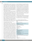
Table 1. Classification of polycythemia and erythrocytosis.
Primary - functional abnormalities expressed in erythroid pro-
genitors
Acquired
Polycythemia vera (JAK2 mutations)
Familial
Primary familial & congenital polycythemia or erythrocytosis (EPOR mutations)
Erythrocytosis due to SH2B3 mutations
Secondary to increased erythropoietin
Acquired
Carboxyhemoglobinemia Erythropoietin doping Erythropoietin-secreting tumor High altitude
Lung or heart disease Smoking
Familial
Left-shifted oxygen dissociation curve 2,3-diphosphoglycerate deficiency High O2 affinity hemoglobins
Methemoglobinemia
Mutations in hypoxia-sensing pathway genes
EGLN1 (PHD2) mutations
EPAS1 (HIF-2α) mutations
VHL mutations (includes Chuvash erythrocytosis), typically homozygous
or compound heterozygous
Gain-of-function mutation of the EPO gene
Secondary to increased exposures other than erythropoietin Acquired
Cobalt
Insulin growth factor 1
Manganese
Post-renal transplant (increased angiotensin signaling) Testosterone
654
haematologica | 2019; 104(4)
