
Page 197 - 2019_04-Haematologica-web
P. 197
Anti-β2GPI-D5 are not thrombogenic in animals
ABC
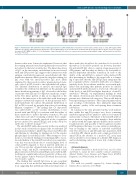
Figure 6. Anti-domain 5 (D5) antibodies interact with β2glycoprotein I (β2GPI) in fluid phase. Reactivity of anti-D5 (aD5), anti-D1 (aD1), or anti-β2GPI-negative (NHS) antibodies (50 μg/mL) against purified β2GPI directly coated on ELISA plates, measured after their incubation with (A) 50 mg/mL, (B) 100 mg/mL, and (C) 200 mg/mL of purified β2GPI ( ) or BSA ( ) in fluid phase. Optical Density (OD) values are expressed as median and interquartile range, and presented as box plots. *P<0.05; **P<0.01.
drawn on this issue. It must be emphasized, however, that the staining intensity varied among different sera and was not related to the level of antibodies. The linear deposition of IgG on the mesenteric endothelium from rats treated with anti-D5-positive IgG suggests their interaction with antigens constitutively expressed on endothelial cells. This distribution pattern differs from the irregular staining for IgG seen with the anti-D1-positive IgG most likely explained by their reaction with a plasma-derived mole- cule, such as β2GPI, bound to the endothelial cell surface. The different distribution of anti-D1 and anti-D5 IgG resembles the well-known difference in the granular and linear distribution patterns of IgG observed in the kidney of patients with SLE and Goodpasture syndrome, respec- tively. The linear pattern of IgG in Goodpasture is the result of the interaction of the antibodies with their target antigen constitutively expressed on the glomerular base- ment membrane. In contrast, the granular distribution of IgG in SLE is caused by irregular deposition of circulating immune complexes.38,39 The finding that C3 deposition was undetectable on the vascular endothelium of rats treated with anti-D5 IgG is consistent with the failure of these antibodies to induce thrombus formation. We and others have provided convincing evidence that comple- ment activation is critically involved in the coagulation process induced by anti-β2GPI IgG and in this study by antibodies to the D1 domain.26,27,40-43
The anti-D4/D5 antibodies present in the sera analyzed in this study selectively recognized the recombinant D5 domain and are likely to inhibit deposition of β2GPI on the endothelium by shielding its binding site for the anionic phospholipid on endothelial cells.44 Our attempt to docu- ment ex vivo reduced binding of circulating β2GPI to vascu- lar endothelium of the anti-D5-treated rats was unsatisfac- tory; this was most likely due to a much higher level of serum β2GPI compared to that of injected antibodies in vivo. The in vitro data obtained under more controlled con- ditions of IgG and β2GPI concentrations showed a fluid phase interaction between anti-D5 IgG and soluble β2GPI, resulting in a significantly reduced reactivity of these anti- bodies against surface-bound β2GPI (when the molecule was bound to a plate).
The finding that anti-D5 IgG have no pro-coagulant effect in our in vivo model has important clinical implica- tions suggesting that individuals with isolated presence of
these antibodies should not be considered to be at risk of thrombosis. It should be pointed out, however, that anti- D1 and anti-D5 IgG often co-exist in a large proportion of APS patients, and that they are likely to be susceptible to anti-D1-dependent thrombus formation. In view of the ability of the anti-D5 IgG to interact with soluble β2GPI, thus preventing its binding to the target cells, it is tempt- ing to speculate that the anti-D5 IgG may antagonize the pro-coagulant activity of anti-D1 antibodies, according to antibody levels. In accordance with this, we recently pub- lished data indicating that patients positive for anti-D1 and anti-D4/5 antibodies have a lower risk of thrombosis if the levels of anti-D4/5 are higher than those of anti-D1 antibodies.7,9 Overall, our experimental findings fit with the clinical observation and offer new tools for stratifying patients into different risk categories. This would help in better preventing recurrences of the clinical manifestations and avoiding overtreatment, thus ultimately improving the patients’ quality of life and sparing them treatment side-effects.
In conclusion, the data presented in this work indicate that, unlike the anti-D1 positive sera, those containing antibodies against D5 are unable to induce clot formation and vascular occlusion. The failure of the anti-D5 antibod- ies to promote coagulation is due mainly to their inability to interact with the target epitopes hidden on the surface- bound molecule, and possibly to the recognition of native β2GPI in plasma that may, to some extent, potentially pre- vent its binding to the surface of activated endothelial cells. The detection of anti-D5 antibodies in patients with a doubtful APS clinical profile and a single positivity for anti-β2GPI in the absence of a positive aCL assay may offer a valuable tool for ruling out a definite APS diagnosis and for identifying subjects at lower risk of clinical mani- festations.
Acknowledgments
The authors would like to thank Linda Vuch, Luca De Maso and Paola A. Lonati for their valuable technical contribution; Michael Mahler, Gary Norman and Filippo Sarra (INOVA Diagnostics and Werfen Italia) for their support.
Funding
This work was partially supported by Istituto Auxologico Italiano, Ricerca Corrente 2016 (PLM).
haematologica | 2019; 104(4)
825
