
Page 25 - 2019_02-Haematologica-web
P. 25
CD30+ primary cutaneous LPD
(CD158k) by neoplastic cells, a potential target reviewed in the next section.47
Altered expression of the T-cell receptor/CD3 complex, T-cell receptor-associated transcription factors and signal transduction molecules is a common feature of systemic and cutaneous CD30+ lymphoproliferations, a finding that may be of potential diagnostic utility.48
Monoclonal rearrangement of the TCR gene occurs in 65% to 90% of cases of pcALCL in contrast with a much lower frequency in LyP types A and B.12,49
Genetic variants of primary cutaneous anaplastic large cell lymphoma
pcALCL seems to carry the same molecular alterations as systemic ALCL, but with dramatic differences in frequen- cies. From the molecular point of view, most pcALCL cases would currently be classified as triple-negative (ALK-, DUSP22-, TP63-negative), since they do not carry any of these precise underlying molecular alterations. However, these lymphomas have an entirely different prognosis from their systemic counterparts, with some of them
showing spontaneous regression and an absence of pro- gression, with long-term survival in the vast majority of the cases (Figure 1). The molecular alterations present in pcALCL are, in descending order of frequency: (i) DUSP22 rearrangements (Figure 2);11,50-61 (ii) ALK translocations (Figure 3);62-65 (iii) TP63 rearrangements;27,66 and (iv) NPM1- TYK2 gene fusion.67-69 ALK translocations are much less frequent than in systemic lymphomas, having been described in isolated cases or very small series.63-65 TP63 rearrangements and NPM1-TYK2 gene fusion are excep- tional and only isolated cases have been reported.27,67-69
ALK-negative primary cutaneous anaplastic large cell lymphoma with DUSP22 rearrangements
DUSP22 rearrangements in systemic ALK– ALCL have been associated with a better prognosis, similar to that of ALK+ systemic ALCL, compared with TP63-translocated or triple-negative cases.50,51 About 20% of pcALCL cases harbor the DUSP22-IRF4 translocation, with no correla- tion with MUM1/IRF4 protein expression.11,52,53 First reported in LyP, a particular biphasic histological pattern has been linked to the presence of DUSP22 rearrange- ments in pcALCL (Figure 2). This pattern is characterized
A
BC
AB
C
E
D
F
D
E
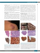
Figure 2. ALK-negative primary cutaneous anaplastic large cell lymphoma with DUSP22 rearrangements. (A) Clinical picture of a large ulcerated tumor in the left malar region of an elderly woman showing (B) the typical CD30 staining of a case of pcALCL with DUSP22 rearrangement (Ref. 58, courtesy of Onaindia et al.), evidencing extensive epidermotropism with CD30+ small-to-medium- sized T-lymphocytes that simulate pagetoid reticulosis lesions. (C) Hematoxylin & eosin-stained panoramic view of the case in (E) and (F) showing the tumor nodule with profuse dermal involvement. (D) Detail, stained with hematoxylin & eosin, of another case exemplifying the biphasic epidermal and dermal lympho- cytic infiltrate. (E,F) (x40), CD3 stain highlights the intraepidermic neoplastic T cells in another case with DUSP22 rearrangement.
Figure 1. “Classic”, ALK-, DUSP22-, TP63- (triple negative) primary cutaneous anaplastic large cell lymphoma. (A) Clinical picture showing two adjacent tumoral erythematous nodules located in the scapular region simulating der- matofibrosarcoma protuberans. (B,C) (x40), Hematoxylin & eosin stain showing the dermal infiltration consisting of a circumscribed infiltrate, composed of arranged large lymphoid cells with absent or subtle epidermotropism. (D,E) (x40), CD30 stain showing positivity in the membrane and Golgi of the tumoral large cells.
haematologica | 2019; 104(2)
229
