
Page 24 - 2019_02-Haematologica-web
P. 24
L. Prieto-Torres et al.
a formalin-resistant epitope (Ber-TRAF1A). They found strong TRAF1 expression in the tumor cells in most LyP cases, in contrast to tumor cells of primary and secondary pcALCL, in which TRAF1 expression was much more restricted.21 Benner et al. also studied this marker in con- junction with MUM1, Bcl2 and CD15, but found it to be of no prognostic or diagnostic utility.22 We review the role of apoptosis in LyP pathogenesis in greater depth in the section on pcALCL, below.
Mahtas et al. studied gene deregulation and spatial genome reorganization near the ALCL translocation breakpoint in ALCL. They described the aberrant expres- sion of Fra2 and of Id2 in LyP, the latter being consistent with the findings of Cotta et al.23,24 They found a lower level of expression in LyP compared with systemic ALCL and argued that gene dosage could be involved in the inva- siveness and progression of LyP.23
P53 mutations have rarely been found in LyP. Kapur et al. reported two cases of LyP with P53 mutations from an analysis of 11 exons of the P53 gene. They found P53 mutations to be infrequent in cutaneous CD30+ lympho- proliferative disorders, and the two patients with LyP who harbored the mutation did not show any changes in the clinical behavior of the disease.25
Notch1 expression has been identified in LyP tumor cells, in which it is associated with the expression of the Notch1 ligand Jagged1, but not of the Delta1 ligand, which was expressed at low or negligible levels.26 The lev- els of expression of Notch1 and Jagged1 were higher in pcALCL samples, a finding that is discussed in greater detail in the section on pcALCL, below.
To date, no ALK fusions have been reported in LyP, in contrast to pcALCL, and no TP63 rearrangements have been found in these patients.25,27,28
Primary cutaneous anaplastic large cell
lymphoma
pcALCL is, by definition, a CD30+ large T-cell neoplasm composed of large cells with an anaplastic, pleomorphic or immunoblastic morphology. The CD30 antigen is expressed in more than 75% of tumor cells. pcALCL resembles other forms of ALCL but arises primarily in the skin.1 The clinical course of pcALCL differs from that of systemic forms of ALCL, both ALK+ and ALK-, which explains why it has been classified as a distinct category.29 However, there is some overlap between systemic and primary cutaneous forms of ALCL, whereby they share some molecular alterations, suggesting that other genetic and biological differences are likely to exist and therefore need to be identified.30
Primary cutaneous large T-cell lymphoma may also be the result of MF tumor progression. Thus, in patients with pcALCL, a current or previous diagnosis of MF must be excluded. The differential diagnosis between pcALCL and transformed CD30+ MF may be challenging, except when there is a clinical presentation with a previous or simulta- neous patch-plaque stage MF lesion. Genetic differences between pcALCL and transformed MF have been found using array-based comparative genomic hybridization.31 From a clinical point of view, patients with pcALCL typi- cally present with solitary or localized nodules or tumors or, more unusually, papules, with frequent ulceration and rapid evolution in some cases that may simulate aggres- sive lymphomas. The presence of multiple lesions in 20%
of cases can hinder the differential diagnosis with type C LyP, which features borderline lesions.7 This cutaneous lymphoma occurs predominantly in males (male:female ratio 3-2:1). It is more frequent in people in their sixth decade, but may also appear in childhood.32 It has been reported that pcALCL is a common form of cutaneous lymphoma in immunosuppressed patients, such as indi- viduals infected with human immunodeficiency virus and organ transplant recipients.33,34 However, in contrast to what usually happens in patients with B-cell lymphomas with CD30+ large cells, especially in immunosuppressed patients, expression of Epstein-Barr virus by the tumor cells is extremely rare or absent in pcALCL with the T/null cell phenotype.32 When it does appear, it is essential to rule out a diagnosis of a B-cell lymphoma with T-cell markers and CD30 expression, such as plasmablastic lymphoma or primary effusion lymphoma.
There is extracutaneous involvement in about 10% of cases, usually with infiltration of locoregional lymph nodes. In these cases it is important to establish the sequence of presentation in order to rule out cutaneous involvement by systemic ALCL, which has an entirely dif- ferent prognosis. Characteristically, locoregional lymph node involvement is not related to bad prognosis in pcALCL. Clinical presentation with extensive skin lesions on legs or arms is the only risk factor associated with a sta- tistically significantly worse prognosis.7,35,36
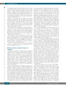
The classic histological pattern described in pcALCL consists of a circumscribed nodular infiltrate that is mostly dermal, composed of arranged large lymphoid cells and usually with absent or subtle epidermotropism5 (Figure 1). However, several variants of this histological pattern have been described, some of which are related to molecular findings described later in this review.5 Neutrophils and eosinophils are usually scattered in classic forms, although rich variants, which are usually more common in immunosuppressed patients, have been reported. The presence of this rich granulocytic infiltrate may be explained by the release of interleukin-8, whose levels are elevated in cultured tumor cells and in the serum of these patients.37 Other unusual histological presentations of pcALCL have been reported, including angiocentric or angiodestructive forms,38,39 subcutaneous and keratoacan- thoma-like forms,40 sarcomatoid variants with prominent spindle-cell morphology,41 small cell variants that are rarer than in systemic forms of ALK+ ALCL,42 and an intravascu- lar ALCL that may involve the skin and must be distin- guished from the more common intralymphatic spread of tumor cells in pcALCL, which seems to have no prognos- tic implications.43,44
pcALCL cells usually carry an activated T-cell pheno- type in which CD30 is expressed in more than 75% of tumor cells (the hallmark of the disease) (Figure 1), fre- quently accompanied by CD3, CD4 and CD45RO expression and varying degrees of loss of CD5 and CD2. The expression of other activation markers, such as CD71, HLA-DR and CD25 (IL-2R), has been noted in approximately half of the cases. Cytotoxic markers (TIA1, perforin and granzyme B) and cutaneous lympho- cyte antigen (CLA, HECA-452) may also be found in around half of the patients.45 In contrast to systemic ALK– ALCL, in which approximately 43% of cases are EMA+,46 EMA is often negative or only focally positive in pcALCL. Interestingly, pcALCL shares with transformed MF and Sézary syndrome the expression of KIR3DL2
228
haematologica | 2019; 104(2)
