
Page 148 - 2019_02-Haematologica-web
P. 148
C. Hoareau-Aveilla et al.
cantly decreased methylation levels of CpGs in NPM- ALK+ cancer cells (Online Supplementary Figure S2C). Collectively, these data suggest that the silencing of both miR-195 and miR-497 is due, at least in part, to NPM-ALK expression and aberrant MIR497HG promoter methyla- tion in ALCL cells.
Forced expression of miR-497 suppresses cell cycle progression and prevents the growth of NPM-ALK+ ALCL cells
To investigate possible biological functions (potential effects on cell proliferation) of miR-195 and miR-497 in NPM-ALK+ cells, the two miRNAs were ectopically
AB
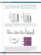
Figure 3. Overexpression of miR-497 reduces in vitro viability and growth of NPM-anaplastic lymphoma kinase (ALK)-positive(+) lymphoma cells. Cell viability deter- mined by MTS assay of NPM-ALK+ lymphoma COST, KARPAS-299 (KARPAS) and SU-DHL1 cells 72 hours (h) after transfection with mimic miR-195 (miR-195) (A) or mimic miR-497 (miR-497) (B). Data represent means±Standard Error of Mean (bars) from 3 independent biological replicates, ***P<0.0001; unpaired two-tailed Student’s t-test with Welch’s correction.
Figure 4. Ectopic expression of miR-497 induces cell cycle arrest and reduces growth in vivo of NPM-anaplastic lymphoma kinase (ALK)-positive(+) lymphoma cells. NPM-ALK+ ALCL COST, KARPAS-299 (KARPAS) and SU-DHL1 cells were transfected with negative control miRNA (miR-CTL) or mimic miR-497 (miR-497). Cell cycle was analyzed (A) and caspase 3/7 activity was measured (B). Data were normalized against the miR-CTL condition. Each experiment was repeated 3 times. (C) NPM- ALK+ COST, KARPAS-299 (KARPAS) and SU-DHL1 cells transfected either with miR-CTL or miR-497 were injected subcutaneously in the left or right flank of immuno- deficient mice, respectively (n=6). Tumor volume was evaluated over time by caliper measurements. Data represent mean±Standard Error of Mean, *P<0.05; **P<0.001; ***P<0.0001; ns: not significant; unpaired two-tailed Student’s t-test with Welch’s correction. (D) Micrographs of hematoxylin & eosin staining of excised miR-CTL or miR-497 tumors (original magnification x40). Arrows indicate cells with phenotypical hallmarks of cellular degeneration.
AB
CD
352
haematologica | 2019; 104(2)
