
Page 147 - 2019_02-Haematologica-web
P. 147
CDK4/CDK6 inhibitors in ALK-positive lymphomas
ABC
DEF
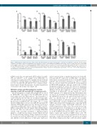
Figure 2. NPM-anaplastic lymphoma kinase (ALK) activity and DNA methylation mediate downregulation of miR-195 and miR-497. Quantitative RT-PCR analysis of miR-195 and miR-497 expression in NPM-ALK-positive lymphoma cell lines, COST, KARPAS-299 (KARPAS) and SU-DHL1, transfected with either an irrelevant siRNA as the negative control (si-CTL) or a siRNA targeting ALK mRNA (si-ALK) (A and D) or treated for 72 hours (h) or not (PBS) with crizotinib (B and E) or treated for 96 h or not (PBS) with decitabine (C and F). SNORD44 was used as an internal control and the relative ratio of miR-195 and miR-497 expression was expressed as 2– ΔΔCt relative to untreated cells. Data represent means±Standard Error of Mean (bars) from 3 independent experiments, **P<0.01; ***P<0.0001; unpaired two- tailed Student’s t-test with Welch’s correction.
miRNA array data, we performed qPCR analysis for miR- 195 and miR-497 on 23 samples from NPM-ALK+ by selecting cases with at least 50% lymph node involvement compared to 12 RLNs (Figure 1B). Significant downregula- tion of both miR-195 and miR-497 was confirmed in pri- mary biopsies despite the presence of a mixed population of neoplastic and normal cells (Figure 1B).
NPM-ALK activity and DNA methylation mediate silencing of miR-195 and miR-497 in lymphoma cells
The downregulation of miR-195 and miR-497 observed in NPM-ALK+ cells suggests that the NPM-ALK+ protein itself might drive this phenomenon. This hypothesis is corroborated by the fact that, in our initial screen, miR- 195 and miR-497 were up-regulated following NPM-ALK knockdown in KARPAS-299 cells (Figure 1A). To verify whether NPM-ALK is involved in miR-195 and miR-497 silencing, NPM-ALK expression was knocked down in 2 other human NPM-ALK+ ALCL cell lines (COST and SU- DHL1) using siRNA directed against ALK mRNA (Figure 2A and D). This was followed by reducing NPM-ALK activity with crizotinib treatment, a pharmacological inhibitor of ALK tyrosine kinase activity (Figure 2B and E). Western blotting analysis (Online Supplementary Figure S1A and B) showed that NPM-ALK was down-regulated and inhibited. Both inhibition of ALK gene expression
and its activity induce a significant increase in both miR- 195 and miR-497 expression in NPM-ALK+ cells (Figure 2A-E). Next, we measured the levels of both miR-195 and miR-497 after decitabine treatment in order to confirm that the observed miR-195 and miR-497 downregulation in NPM-ALK+ lymphoma cells was, indeed, due to DNA methylation (Figure 2C and F). We detected a significant increase in miR-195 and miR-497 expression in DNMT inhibitor-treated cells compared to the drug vehicle alone (DMSO) in all NPM-ALK+ cell lines (Figure 2C and F). STAT3 appears to be implicated in this mechanism (data not shown). To determine the DNA methylation status of the MIR497HG promoter, the miR-195 and miR-497 host gene on chromosome 17, we carried out bisulfite sequencing analysis of genomic DNA isolated from the three NPM-ALK+ cell lines: KARPAS-299, SU-DHL1 and COST and normal CD4 T lymphocytes expressing CD30 antigen activated using CD3/CD28 antibodies (n=3) (Online Supplementary Figure S2A). The results showed sig- nificantly high levels of methylation of all CpGs located in the region upstream of miR-195 and miR-497 promot- ers in the KARPAS-299 and SU-DHL1, and a few CpGs (CpG1, CpG2 and CpG3) in COST tumor cells. We observed lower levels of CpG methylation in the region upstream of miR-195 and miR-497 promoters in normal cells (Online Supplementary Figure S2B). Decitabine signifi-
haematologica | 2019; 104(2)
351
