
Page 117 - 2019_02-Haematologica-web
P. 117
ASNase hypersensitivities are IgG/IgE dependent
rily detected in cells obtained from the liver (73.6%), blood (11.4%), and spleen (5.0%) 8 h after administration (Online Supplementary Figure S1A). Among CD45+ leuko- cytes, ASNase accumulates primarily in macrophages/monocytes of the liver and blood (95.1%, and 93.7%, respectively; Online Supplementary Figure S1A and B), to a lesser extent in neutrophils (1.1% and 2.1%, respectively), but not in other immune cells. Supporting the role of macrophages in ASNase clearance, the uptake of ASNase was assessed using a murine macrophage cell line (i.e., RAW 264.7 cells). ASNase positive RAW cells were detected after 5 minutes of incubation with ASNase
and the percentage of ASNase+ RAW cells plateaued after about 2 h of incubation with the drug (Online Supplementary Figure S2A-B). Therefore, it is likely that the binding of ASNase to naïve macrophages/monocytes is associated with drug clearance rather than antigen-specific recognition.
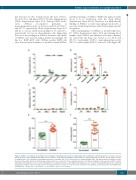
After sensitizing mice to ASNase, as described previous- ly21 (Online Supplementary Figure S3A) and staining blood samples with ASNase and immune cell surface markers, we found that the drug was bound ex vivo by B cells (53.3%), neutrophils (14.5%), macrophages/monocytes (32.9%), and basophils (69.5%), but not T cells (Figure 1B).
AB
CD
EF
Figure 1. ASNase is recognized ex vivo by B cells, neutrophils, macrophages/monocytes, and basophils after sensitization. (A) Peripheral blood cells were collected from naïve mice, cultured with labeled ASNase for 30 minutes, and analyzed for ASNase positive immune cells by flow cytometry. (B) The ex vivo ASNase-specific recognition by B cells, neutrophils, macrophages/monocytes, basophils, and T cells of the blood were analyzed within CD45+ populations by flow cytometry of sensi- tized (red data points) and non-sensitized (green data points) mice on Day 23 of the sensitization protocol. (C) ASNase binding to total leukocytes (CD45+) and (D) anti-ASNase IgG antibodies were measured throughout the sensitization protocol. Anti-ASNase IgE antibodies were measured by (E) ELISA and (F) flow cytometry using plasma samples collected on Day 23 of the sensitization protocol. A total of 5 to 20 mice were included in each analysis, as indicated, and P value significance is indicated as * for P<0.05, ** for P<0.01, *** for P<1x10⁻³, and **** for P<1x10⁻⁴.
haematologica | 2019; 104(2)
321
