
Page 26 - 2019_01-Haematologica-web
P. 26
U. Oyarbide et al.
addition to severe anemia, individuals with DBA may dis- play physical anomalies that include thumb, upper limb, craniofacial, cardiovascular and kidney malformations, and short stature. DBA patients have a 25% higher risk of developing myelodysplastic syndromes (MDS), acute myeloid leukemia (AML), and osteosarcoma.
Diamond-Blackfan anemia is an autosomal dominant disorder with a disease incidence of 5-7 per million live births, equally distributed between genders.28 DBA patients have mutations in approximately 20 genes encod- ing ribosomal proteins; the most common (25%) is RPS19.29 Frameshift, splice defects, intragenic deletions and insertions, nonsense, as well as missense mutations have all been identified. Mutations involve other riboso- mal genes: RPL5 (7%), RPL26 (6.6%), RPL11 (5%), RPS10 (3%), RPS26 (3%), RPL35A (3%), RPS24 (2.4%), RPS17 (1%), RPL15, RPS28, RPS29, RPS7, RPS15, RPS27a, RPS, RPL9, RPL18, RPL26, RPL27, RPL31.3 These findings sup- port DBA as a disorder of ribosomal biogenesis and/or function. Mutations in three non-ribosomal proteins, GATA1, TSR2, and EPO, are also associated to DBA.3,29 It is hypothesized that DBA results from apoptosis due to aberrant activation of TP53 that induces cell cycle arrest or apoptosis in response to ribosomal stress.30 Some of the reports implicating TP53, as reviewed below, are based on MO-mediated effects.
In two different studies, rps19-deficient zebrafish were
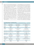
Table 3. Comparison of human, mouse and zebrafish blood systems. Human
created using MO. Knockdown of rps19 in zebrafish reca- pitulates the hematopoietic and developmental pheno- types of DBA, including erythropoietic failure with severe anemia, with cell cycle arrest and increased apoptosis, with p53 upregulation. The rps19-deficient phenotype was rescued by injection of zebrafish rps19 mRNA.31-33 Moreover, these phenotypes were not rescued by express- ing rps19 mRNAs with a missense or nonsense mutation found in DBA patients.32 Co-injection of MOs against rps19 and p53, showed a complete rescue of the morpho- logical abnormalities, but did not rescue the hematologic defects. These results suggest that there is an erythroid specificity in Rps19 deficiency in zebrafish, independently of Tp53 activity. (See below for further discussion on Tp53 in DBA pathogenesis).34
Chakraborty et al. analyzed the effect of MO-mediated loss of rpl11 in zebrafish. Knockdown of rpl11 led to mor- phological defects in the developing brain, head, and eyes, and pericardial edema. These phenotypes appear specific as the investigators were able to suppress the morphant by co-injection of MO-resistant rpl11 mRNA. Similar to the loss of Rsp19 function, knockdown of rpl11 resulted in an upregulation of tp53 and mdm2. Moreover, co-injection of rpl11 and tp53 MO rescued the developmental defects and reduced apoptosis, suggesting that ribosomal dys- function due to the loss of Rpl11 activates a Tp53-depen- dent response to prevent faulty embryonic development.
Adult HSC
Blood cell types
Erythrocytes (life span)
Platelets
(life span)
Neutrophils (life span)
Primitive myelopoiesis
Definitive myelopoiesis
Primitive erythropoiesis
Definitive erythopoiesis
Circulation
Primitive thrombopoiesis
Definitive thrombopoiesis
Developmental HSC
B cells T-cell
maturation
Mouse
Bone marrow
Erythrocytes, granulocytes,
lymphocytes and platelets
Without nucleus (60 days)
Platelets
(4 days)
Twisted toroid with a central hole mpo expressing cell
(12.5 hours)
Yolk sac (E7.25-E10), AGM, fetal liver
(after E9.5)
Fetal liver (E9.5) Bone marrow
Yolk sac
(E7.0)
Yolk sac (E9.5),
fetal liver (E12.5) and then bone marrow
Begins at E8.5
N/A
Bone marrow
AGM next fetal liver and finally bone marrow
Bone marrow Thymus
(E10-12)
Zebrafish
Kidney marrow
Erythrocytes, granulocytes, lymphocytes
and thrombocytes
With nucleus (at least 10 days)
Thrombocytes
(4 days)
Segmented nuclei with
two or three lobes
mpo expressing cells (3.5 days)
ALM (~11 hpf) and CHT (~24 hpf)
Kidney
(~HSC starts seeding at 4 dpf)
ICM
(~12 hpf)
CHT (2-6 dpf) and then kidney marrow (4 dpf)
Begins at 24 hpf
CHT (~48 hpf)
Kidney marrow (~5 dpf)
AGM next CHT and finally kidney marrow
Kidney marrow Thymus
(7 dpf)
Bone marrow
Erythrocytes, granulocytes,
lymphocytes and platelets
Without nucleus (115 days)
Platelets
(8-9 days)
Segmented nuclei with up to four lobes mpo-expressing cells (5.4 days)
Yolk sac, AGM, fetal liver
Fetal liver and bone marrow
Yolk sac
(3-4 weeks)
Yolk sac (4 weeks) Fetal liver (5-6 weeks) and then bone marrow
Begins at 8 weeks
N/A
Bone marrow
AGM next fetal liver and finally bone marrow
Bone marrow Thymus
(8-9 weeks)
AGM: aorta-gonadal-mesonephros; ALM: anterior lateral mesoderm; CHT: caudal hematopoietic tissue; dpf: days post fertilization; ICM: intermediate cell mass.
16
haematologica | 2019; 104(1)
