
Page 24 - 2019_01-Haematologica-web
P. 24
U. Oyarbide et al.
SDS is emerging as a disorder in proteostasis and ribo- some maturation (Table 1).4 The molecular basis for how these phenotypically and genotypically heterogeneous conditions result in single or multiple cytopenias remains poorly understood. No common pathway has yet been established, but zebrafish studies have suggested TP53 responses. Activation of the TP53 pathway in mediating marrow failure has been reported for DC,5 FA,6 and a novel bone marrow failure syndrome.7 The TP53 path- way has been suggested to mediate marrow failure for other inherited neutropenias such as SCN and SDS.8 Environmental exposures can accelerate marrow failure, for example, aldehydes producing DNA crosslinks in FA.9 How epigenetics and genetic co-modifiers contribute to these diseases is even less understood. Investigating the molecular basis of the IBMFS will lead to a greater under- standing of hematopoiesis, and development and mainte- nance of non-hematologic tissues. Since the IBMFS consti- tute leukemia or cancer predisposition syndromes, insights into their pathophysiology will also benefit our understanding, prevention, and perhaps treatment of can- cer and age-related genetic changes.
Zebrafish model to study inherited bone marrow failure syndromes
Zebrafish (Danio rerio) have gained popularity as a model organism for a number of reasons. Approximately 70% of all human genes have a zebrafish ortholog.10 Genes are orthologs if they evolved from a common gene, and orthologs typically share similar function. (The
Human Genome Organization has adopted a nomencla- ture for gene and protein expression among different species, which we show using SDS as an example in Table 2.) In addition to lower maintenance and breeding costs, zebrafish provide major advantages to mice: their large clutch size of externally fertilized eggs, transparent embryos, quicker development (all major organs develop and begin functioning during the first 5 days), and short generational time to gamete formation.11 A high degree of genetic and morphological similarity in hematopoiesis between zebrafish and humans suggests that zebrafish can provide valuable insights into the pathogenesis of IBMFS. Developmental hematopoiesis in the zebrafish is comparable to that observed in mice or humans (Figure 1).12-15 One notable difference is that the site of definitive hematopoiesis lies in the zebrafish kidney perivascular space, not the bone marrow. Since the hematopoietic stem cell (HSC) niche provides protection and regulation of self-renewal and differentiation of HSC into blood cells, this difference may be important in non-cell autonomous processes.
Studies using zebrafish have facilitated our understand- ing of vertebrate hematopoiesis and aberrant hematopoiesis in diseases. Hematopoietic and non- hematopoietic lineage-specific transgenic reporter strains are available. They have been useful for the identification and characterization of genes for embryonic hematopoiesis, erythropoiesis, and modeling of human blood diseases (Table 3).16-19 In addition to a collection of zebrafish mutants induced by N-ethyl-N-nitrosourea or
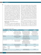
Table 1. Inherited bone marrow failure syndromes. Disease Prevalence Male-to-female
per 1,000,000 ratio
Diamond-Blackfan 5-7 1:1 anemia (DBA)
Dyskeratosis 1 3:1 congenita (DC)
Fanconi anemia (FA) 3 1.2:1
Shwachman-Diamond 13 1.7:1 syndrome (SDS)
Congenital Unknown amegakaryocytic (less than thrombocytopenia 100 cases (CAMT) reported)
Severe congenital 5
neutropenia (SCN)
Symptoms
Genes involved and their estimated frequency
RPS19 (25%), RPL5 (7%), RPS26 (6.6), RPL11 (5%), RPL35a (3%), RPS10 (3%), RPS24 (2.4%), RPS17 (1%), RPL15, RPS28, RPS29, RPS7, RPS15, RPS27a, RPS27, RPL9, RPL18, RPL26, RPL27, RPL31, TSR2, GATA1, EPO
DKC1 (17-36%), TERC (6-10%), TERT(1-7%), NHP2 (<1%), NOP10 (<1%), CTC1 (1-3%), WRAP53 (3%) and TINF2 (11-24%), ACD, PARN, RTEL1, USB1, TCAB1, POT1, TPP1, WRD79, TR, NOLA2, NOLA3
FANCA (65%), FANCB (<1%),
FANCC (14%), FANCG (10%), FANCD1/BRCA2 (<1%), FANCD2 (<1%), FANCE (4%), FANCF (4%), RAD51, FANCC1, FANL, FANCL, FANC, PALPB2, RADC51C, SLX4, FANCQ. BRCA1, FANCT
SBDS (90%) DNAJC21 EFL1, SRP54
MPL
ELANE, GFI1, HAX1, G6PC3, VPS45, JAG1, CSF3R, WAS, SRP54
Cancer predisposition
AML, MDS, ALL, Hodgkin and non-Hodgkin lymphomas, osteogenic sarcoma, breast cancer, hepatocellular carcinoma, melanoma, fibrohistiocytoma,
gastric cancer, colon cancer
AML, solid tumors
AML, solid tumors
AML, MDS
AML, MDS
AML, MDS
Erythroid failure, congenital malformations, growth retardation, short stature. Thumbs, upper limbs, hands, and craniofacial, urogenital, and cardiovascular anomalies are also common
Abnormal skin pigmentation, nail dystrophy, mucosal leukoplakia, pulmonary fibrosis, and bone marrow failure
Developmental abnormalities in a number of organ systems and bone marrow failure
Exocrine pancreatic insufficiency, bone marrow dysfunction and skeletal abnormalities
Thrombocytopenia and megakaryocytopenia
Neutropenia
AML: acute myeloid leukemia; ALL: acute lymphocytic leukemia; MDS: myelodysplastic syndromes.
14
haematologica | 2019; 104(1)
