
Page 196 - 2019_01-Haematologica-web
P. 196
C. Tromeur et al.
fusion-only scintigraphy should be performed using a reduced dose of 99mTc-MAA (approximately a quarter of the usual dose administrated for a one-step V/Q scan). Because of the low frequency of co-morbid pulmonary disorders, PE can be excluded in most cases on the basis of a normal perfusion pattern. Ventilation images should only be per- formed in the case of abnormal perfusion images.
Conclusion
Based on the available data, direct comparisons of safety and efficiency between CTPA and V-Q lung scanning do not seem valid. The available studies are based mostly on techniques that are outdated with regard to the current and presently evolving techniques, for both CTPA and V-Q
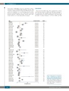
Figure 3. Meta-analysis of non-diagnostic results of ventilation-perfusion lung scan- ning and computed tomography pulmonary angiography in pregnant patients with sus- pected acute pulmonary embolism. The number and type of additional imaging tests are provided in Table 3. V-Q: ventilation-per- fusion; CTPA: computed tomography pul- monary angiography.
186
haematologica | 2019; 104(1)
