
Page 63 - 2018_12-Haematologica-web
P. 63
Progressive hematopoietic defects in ASXL2 KO mice
ABC
DEF
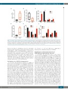
Figure 6. Cell-intrinsic effects of Asxl2 deficiency on lymphoid and myeloid lineages. Lethally irradiated mice transplanted with either wild-type (WT) or Asxl2 knock- out (KO) bone marrow (BM) cells were analyzed after one year. (A) Number of white blood cells (WBCs) in the peripheral blood of recipient mice. (B) Number of thy- mocytes in recipient mice. (C) Proportion of donor-derived (CD45.2+) DN, DP, CD4+SP and CD8+SP cells in thymi of recipient mice. (D) Spleen cellularity. (E) Percentages of CD45.2+ B, T and myeloid cells in the spleen of mice transplanted with either WT or KO cells. (F) Frequencies of erythroid precursors in spleens of recipient mice. Bars represent mean±Standard Error of Mean. *P<0.05, **P<0.01, ***P<0.001, ****P<0.0001.
However, the BM of old KO mice showed a substantial reduction in the frequencies and absolute number of proB (CD43+B220+), preB (CD43–IgM–B220+), immature B (CD43–IgM+B220+), and mature B cells (Figure 5A and B), indicating an age-dependent paucity in B-cell develop- ment. Further fractionation of proB cells using the scheme proposed previously,36 revealed a lower proportion of CD24–BP1– (Fraction A) and CD24+BP1– (Fraction B) proB cells in the old Asxl2 KO mice (Figure 5A and B), demon- strating a partial arrest of B-cell maturation.
Cell-intrinsic role of ASXL2 in hematopoiesis
To test the cell-autonomous function of ASXL2 in hematopoietic development, we transplanted lethally irra- diated mice with either WT or Asxl2 KO BM cells and ana- lyzed the hematopoietic compartment one year later. We observed that the recipient mice transplanted with Asxl2 KO BM cells tended to have higher WBC counts in periph- eral blood (Figure 6A), similar to the age-dependent increase in WBC observed in KO mice (Figure 2A). We also noted decreased thymocyte cellularity and accumula- tion of donor-derived DN cells in the thymi of mice trans- planted with Asxl2 KO BM compared with those trans- planted with WT BM (Figure 6B and C). Spleens of Asxl2 KO BM-recipients were enlarged and exhibited elevated proportion of myeloid cells (Figure 6D and E). Moreover, our analysis also indicated a higher proportion of ery- throid precursors in spleens of mice reconstituted with Asxl2 KO BM cells compared with the WT cells (Figure 6F), similar to the phenotype observed in old Asxl2 KO mice. Overall, the recipient mice recapitulated hematopoi-
etic features of old Asxl2 KO mice, suggesting a hematopoietic cell-intrinsic function of ASXL2.
Identification of altered gene expression in ASXL2-deficient hematopoietic stem cells
To determine the molecular basis of the defects observed in Asxl2 KO HSCs, we performed global gene expression profiling (RNA-Sequencing) of sorted LSK cells from old and young WT and Asxl2 KO mice. Comparison of transcriptomic profiles of LSK cells from old WT and Asxl2 KO identified more than 2500 genes differentially expressed (983 genes up-regulated and 1653 genes down- regulated in the KO cells; FDR<0.1) (Online Supplementary Table S6), including those involved in myeloid differentia- tion such as Csf1, Gfi1b, Gata2, Hoxa9, Hoxa5, Mpl, Cdk6, Ccr1 and Ets1 (Online Supplementary Figure S15A). Asxl2-deficient LSK cells from old mice exhibited more pronounced changes in gene expression compared with the young mice (Figure 7A and B and Online Supplementary Figure S15B). However, a significant overlap of genes com- monly up-regulated and down-regulated in the KO LSK cells was noted within the two age groups (Figure 7C and Online Supplementary Table S6). Interestingly, GSEA analy- sis revealed that the expression of RUNX1-RUNX1T1 tar- gets37 inversely correlated in the KO cells (Figure 7D and Online Supplementary Figure S15C). Also, genes down-reg- ulated in immature BM progenitors upon silencing of CBFA2T3 (also called ETO2)38 were also suppressed in the LSK cells from both old and young Asxl2 KO mice (Figure 7E and Online Supplementary Figure S15D). CBFA2T3 is involved in the translocation t(16;21)(q24;q22) with
haematologica | 2018; 103(12)
1987
