
Page 59 - 2018_12-Haematologica-web
P. 59
Progressive hematopoietic defects in ASXL2 KO mice
ABC
DEF
GH
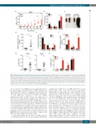
Figure 2 Myeloproliferation and extramedullary hematopoiesis in Asxl2-deficient mice. (A) White blood cell counts in peripheral blood of 8-10 week old wild-type (WT) and Asxl2-deficient mice. Whiskers extend from the minimum to the maximum values. (B) Proportion of B cells (CD19+), T cells (CD3+), and myeloid cells (CD11b+) in the peripheral blood of >1-year old WT and Asxl2 knockout (KO) mice determined using flow cytometry (B and T cells, n=3; myeloid cells, n=11). (C) Representative photograph of spleens isolated from young (11-week old) and old (>1-year old) WT and Asxl2 KO mice. (D) Total leukocyte counts in spleens from >1-year old mice. (E and F) Frequencies (E) and absolute numbers (F) of lymphoid and myeloid cells in spleens of old WT and KO mice. Flow-cytometric analysis on splenocytes was similar to (B) (n=12-13). (G) Frequency of LSK cells in spleen and peripheral blood of old WT and KO mice. (H) Proportion of erythroid progenitors detected in flow cytometric analysis (staining with CD71 and TER119 antibodies) of spleens from old WT and KO mice (n=10). Bars represent mean±Standard Error of Mean. *P<0.05, **P<0.01, ***P<0.001, ****P<0.0001, ns: not significant.
ly.24 As reported,24 Asxl2 KO mice had a significantly shorter life span compared with the WT mice (Online Supplementary Figure S4B). We observed a significant reduction in Asxl2 transcript and protein levels in the BM, spleen and thymus of Asxl2 KO mice compared to WT mice (Online Supplementary Figure S5A-F), whereas the expression of Asxl1 was not affected (Online Supplementary Figure S5G-I). We cultured the BM cells from WT and KO mice for two weeks in myeloid differentiation-promoting conditions and observed an increase in the proportion of CD11b+ cells with a concomitant decrease in Lin–Kit+ cells in the cultures from Asxl2-deficient BM, suggesting altered myeloid differentia- tion (Online Supplementary Figure S6A and B). Moreover, in re-plating assays, we observed a sustained ability of Asxl2 KO BM cells to generate myeloid colonies compared with WT cells (Online Supplementary Figure S6C).
To gain initial insights into the effect of ASXL2 deficien- cy on hematopoiesis in vivo, we periodically analyzed the
peripheral blood (PB) from Asxl2 KO and WT mice begin- ning at eight weeks of age. We observed an age-depen- dent increase in white leukocyte count in the PB of Asxl2- deficient mice, while the numbers were unchanged in the WT mice (Figure 2A). This was associated with reduced red blood cell (RBC) counts and hemoglobin in peripheral blood of Asxl2 KO mice with increasing age (Online Supplementary Figure S7). This observation suggested pro- gressive defects in the hematopoietic compartment in Asxl2 KO mice, and therefore we investigated systemati- cally hematopoietic development in >1-year old mice. Flow cytometric analysis of blood leukocytes from these mice showed a higher proportion of myeloid (CD11b+) and a reduced proportion of lymphoid (CD19+ and CD3+) cells in KO mice (Figure 2B and Online Supplementary Figure S8A). Old Asxl2 KO mice exhibited extensive splenomegaly (Figure 2C and D) which was associated with marked myeloproliferation evident by increased fre-
haematologica | 2018; 103(12)
1983
