
Page 54 - 2018_12-Haematologica-web
P. 54
X. Liu et al.
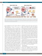
Figure 7. Overview of the alterations in the bone marrow niche of Twist1-deleted mice. Simplified scheme of the normal hematopoietic stem cell (HSC) niche and its alterations in the Twist1-deleted mice. The left panel shows that HSC are around arterioles, sinusoids and endosteum where factors such as C-X-C motif chemokine ligand 12 (CXCL12), vascular cell adhesion molecule1 (VCAM1), stem cell factor (SCF) and osteopontin (OPN) secreted by mesenchymal stem cells (MSC), endothelial cells and osteoblastic cells (OBC) influence their self-renewal, quiescence, retention and differentiation. The right panel summarizes alterations of the niche, HSC and leukemic stem cells (LSC) observed in Twist1-deleted mice.
posing or initiating factors for the evolution of AML and point to TWIST1 as an instructive signal that alters the function of the niche.
The opposing effects of TWIST1 on normal HSC and LSC found in this study are of value. In an effort to eluci- date the underlying mechanism, we performed RNA- sequencing and quantitative real-time polymerase chain reaction analysis. We found that Twist1 deletion leads to increased expression of the Notch ligand Jagged-2 in all the OLC, EC and MSC. LSC from Twist1-deleted chimeric mice have robust expression of all Notch receptors and canonical downstream Notch target genes, suggesting the aberrant activation of Notch signaling. A previous report showed that Notch activation promotes expansion and self-renewal of LSC,40 consistent with our results obtained by deletion of Twist1. We also found activation of Notch receptors and target genes in normal HSC (CD34-LSK) after Twist1 deletion (Online Supplementary Figure S9). In contrast to the promoting role in LSC, Notch activation in HSC has been reported to cause loss of stem cell quies- cence,46 which often correlates with impaired self-renewal capacity of HSC, in line with the observations in our mouse model. Besides the direct impact of activated Notch signaling on LSC and HSC, the augmented prolifer- ation and infiltration of LSC compared to normal HSC could be favorable for their competition for the niche over HSC. Various studies have demonstrated that LSC could positively remodel the BM microenvironment to enhance support of LSC at the expense of HSC,47,48 and this remod- eling may in turn further promote leukemia progression and impair normal hematopoiesis. In addition, the reduced expression of Cxcl12, Scf and Angpt1 in Twist1- deleted mice may also account for the opposing impact of Twist1 deletion on HSC and LSC, since compared with HSC, LSC are less factor-dependent.34,45,49
In consideration of the important role of TWIST1 in reg-
ulating MSC, osteoblasts and EC, and to exclude the inter- ference of hematopoietic cells, which were found to express Twist1 in our previous work,50 we generated the chimeric mouse model, in which Twist1 was diffusely deleted in the BM microenvironment. The BM niche com- prises multiple cell types, which not only closely connect but also communicate with each other via cell factors and adhesion molecules throughout the BM. Due to the com- plexity of the niche, an overall environmental knockout strategy will facilitate the detection of direct and indirect effects of TWIST1 on the niche components. Utilizing our model, we uncovered extensive cellular and factor alter- ations in the BM niche and the AML-like microenviron- mental phenotype resulting from Twist1 deficiency, and demonstrated the essential role of TWIST1 in HSC main- tenance and suppression of AML evolution. To refine the contribution of different cell populations, studies in which Twist1 is modified in specific stromal cell subsets are ongo- ing in our laboratory.
In conclusion, we used a Twist1-deficient chimera model to obtain, for the first time in vivo, direct evidence that TWIST1 in the microenvironment plays a key role in main- taining the hematopoietic phenotype and hampering leukemia progression. These findings provide new insights into the importance of the BM niche for AML develop- ment, and lay the foundation for tackling leukemia from a different angle to improve current treatments.
Acknowledgments
This work was supported by grants from The National Key Research and Development Program of China (2016YFA0100603), CAMS Initiative for Innovative Medicine (2016-I2M-1-017), National Natural Science Foundation of China (81470278, 81670158, 81600138, and 81700106), and Tianjin Municipal Science and Technology Commission grants (17JCZDJC35100 and 17JCQNJC10800).
1978
haematologica | 2018; 103(12)
